

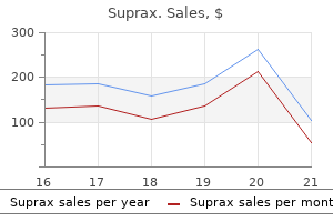
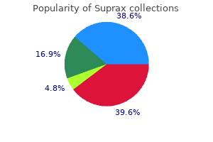
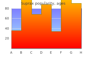
Suprax
Robert O’Connor, MD, MPH
- Professor and Chair, Department of Emergency Medicine, University of
- Virginia, Charlottesville, VA, USA
Suprax dosages: 200 mg, 100 mg
Suprax packs: 30 pills, 60 pills, 90 pills, 120 pills, 180 pills, 270 pills, 360 pills

Cheap suprax 200mg on line
Patients with central stenosis typically have bilateral antibiotic resistant bacteria mrsa purchase suprax 200 mg on-line, non- dermatomal ache involving the buttocks and posterior thighs antibiotic qt prolongation order suprax 100 mg visa. Repeating the bodily examination after fast walking may demonstrate delicate abnormalities bacteria song cheap suprax 200 mg on-line. Progression of cervical and thoracic stenoses may cause myelopathy and paralysis and requires surgical procedure extra usually than lumbar spinal stenosis. Table 7-10 outlines the historic and bodily exam findings associated with the prognosis of spinal stenosis. Patients with typical symptoms who reply to conservative therapy may be managed with out imaging. Physical therapy improves stamina and muscle strength in the legs and trunk; workouts performed with lumbar flexion, such as cycling, may be better tolerated than walking. Epidural corticosteroid injection helps some patients, especially these with radicular ache. Predictors of a positive response to surgical procedure include male gender, youthful age, better walking capability, higher self-rated well being, less comorbidity, and extra pronounced canal stenosis. A recent trial with both a randomized and remark cohort confirmed the next: a. In the intention to treat analysis of the randomized cohort, patients randomized to surgery reported better scores on 1 measure of bodily ache at 2 years than did these randomized to conservative remedy. In the evaluation of the observational cohort, sufferers who chose surgical procedure reported better pain and performance scores than those that selected conservative therapy. Have you crossed a diagnostic threshold for the main hypothesis, spinal stenosis Critical limb ischemia classically presents with ache within the ft at rest that could be relieved by placing the ft in a dependent place. Apply agency stress to the plantar aspect of the great toe for 5 seconds; after releasing the toe, regular shade should return in 5 seconds. Cilostazol a hundred mg twice day by day increases strolling distance by 50% after 3� 6 months of use; pentoxifylline has no effect on strolling distance. Ramipril increases mean ache free walking time by over 200 yards in patients with uncomplicated, steady claudication not already taking an angiotensin-converting enzyme inhibitor or angiotensin receptor blocker. Exercise, especially a supervised train program, can improve walking by up to 150% over 3�12 months. Revascularization, either surgical or percutaneous transluminal angioplasty, is indicated for the next: 1. After attending physical remedy for presumed spinal stenosis for 8 weeks, he reviews some improvement in his exercise tolerance, although he nonetheless has every day pain. An epidural corticosteroid injection supplies extra ache aid, and he is ready to proceed a walking program. Spinal abnormality or intervention (degenerative joint illness, trauma, surgical procedure, drug injection) c. Potential native or systemic source of an infection (skin or soft tissue an infection, endocarditis, osteomyelitis, urinary tract infection, injection drug use, epidural anesthesia, indwelling vascular access) 2. Infection happens by contiguous spread in 33% of circumstances and by hematogenous unfold in 50%. Other organisms embrace Staphylococcus epidermidis, Escherichia coli, Pseudomonas aeruginosa. More widespread in posterior than anterior epidural area, and extra widespread within the thoracolumbar than cervical areas. The most essential predictor of the ultimate neurologic end result is the neurologic standing before surgery, with the postoperative neurologic status being nearly as good as or better than the preoperative status. Antibiotics Vertebral Osteomyelitis Textbook Presentation the basic presentation is unremitting back pain often, however not at all times, with fever. Urinary tract, pores and skin, delicate tissue, vascular entry web site, endocarditis, septic arthritis most commonly found sources, with endocarditis present in one-third of sufferers with vertebral osteomyelitis b. Can also occur as a result of contiguous spread from an adjoining soft tissue infection or direct infection from trauma or surgical procedure. Generally causes bony destruction of two adjacent vertebral bodies and collapse of the intervertebral space. Found within the lumbar spine in 58% of circumstances, thoracic spine in 30%, and cervical spine in 11% b. Complicated by epidural abscess in 17% of cases, by paravertebral abscess in 26%, and disk space abscess in 5% B. C-reactive protein additionally elevated in nearly all sufferers, and may be a greater marker of response to therapy. Culture of a biopsy specimen is positive in about 77% of sufferers (range in research 47�100%). Surgery is necessary provided that neurologic symptoms suggest onset of vertebral collapse inflicting cord compression or growth of spinal epidural abscess; surgery is at all times essential for osteomyelitis related to a spinal implant. Endocarditis must be thought-about in sufferers with both vertebral osteomyelitis or a spinal epidural abscess. Chou R, Qaseem A, Snow V et al; Clinical Efficacy Assessment Subcommittee of the American College of Physicians; American College of Physicians; American Pain Society Low Back Pain Guidelines Panel. Diagnosis and treatment of low again pain: a joint scientific apply guideline from the American College of Physicians, and the American Pain Society. Predictive worth of medical traits in sufferers with suspected cauda equina syndrome. Does the medical examination predict lower extremity peripheral arterial disease Does this older grownup with lower extremity pain have the medical syndrome of lumbar spinal stenosis Office evaluation of backbone and limb pain: spondylotic radiculopathy and other nonstructural mimickers. A is a 24-year-old woman who involves see you because her gums are bleeding when she brushes her teeth. Bleeding due to platelet abnormalities, whether or not because of decreased quantity or abnormal function of platelets, is often small vessel bleeding, and produces such findings as petechiae, bruising, gum bleeding, or nosebleeds. Platelet-related bleeding is mostly not quantitatively vital (ie, platelet-related bleeding tends to not trigger severe blood loss requiring pink cell transfusions). Nonetheless, platelet-related bleeding can nonetheless be clinically essential if a affected person bleeds a small amount into the mind (unusual unless the platelet depend is < 10,000/mcL) or induces an stomach hematoma from vigorous coughing, for instance. Bleeding as a result of coagulation issue abnormalities is extra likely to be quantitatively important, typically occurring in joints, the gastrointestinal tract, mind, retroperitoneum, or at sites of current damage or medical or surgical intervention. Abnormality of the tissue such that minor trauma causes bleeding, corresponding to a toothbrush inflicting gum bleeding from inflammatory gingival disease B. Decreased manufacturing of platelets (1) Medications (examples embrace valproic acid, linezolid, thiazide diuretics, gold compounds, antineoplastic chemotherapy drugs) (2) Bone marrow alternative by malignancy, fibrosis, granulomas (3) Bone marrow aplasia (4) Alcohol (5) B12 deficiency b. She notes that her final menstrual period was considerably heavier than ordinary, and he or she has had an intermittent headache over the last 2 days, partially relieved by acetaminophen. Her oral examination shows evidence of current gingival bleeding, and there are a quantity of palatal petechiae. The second pivotal level is that her historical past additional means that her platelet dysfunction is acquired.
Buy suprax 100 mg online
In the palms of a sophisticated thoracoscopic surgeon best antibiotic for uti least side effects discount suprax 100mg without prescription, further procedures antibiotics not helping uti trusted suprax 100mg, together with diaphragmatic hernia repair antibiotic resistance future order 200mg suprax visa, pulmonary lobectomy, and esophageal atresia/tracheoesophageal fistula repair may additionally be routinely performed in this method. Thoracoscopy presents a variety of advantages in comparability with conventional thoracotomy together with much less pain and higher cosmesis. Moreover, the present decision of high definition optical systems offers better magnification and subsequently superior visualization of the thoracic mediastinum, significantly in the apex. For lobectomy, lung biopsy, and pulmonary decortication, the affected person ought to be within the lateral decubitus position. A 5-mm trocar is gently inserted, and the thorax is insufflated with carbon dioxide to 4 mmHg at a move price of 0. For all these positions, the use of gravity by rotation of the mattress may be helpful to maintain the uninvolved lung and different buildings out of the field. In these situations, a triangular arrangement of the trocars works greatest with the digicam port placed slightly above and between the two working ports. Larger youngsters have wider intercostal areas that can usually accommodate 12-mm trocars for endoscopic linear stapling. Upon completion of the process, all incisions are infiltrated with native anesthetic and closed in a single or two layers utilizing non-absorbable sutures. Typical tube sizes range from 12 to 28 Fr, depending on the scale of the affected person and kind of pleural drainage anticipated. The tube ought to all the time exit the physique anterior to the mid-axillary line extending all the way down to the anterior superior iliac backbone. Heavy non-absorbable suture is used to safe the tube to the skin followed by software of an occlusive, adherent dressing. Experience with a total muscle-sparing strategy for thoracotomies in neonates, infants, and children. Thomas Gibson in 1697 precisely described the clinical options of esophageal atresia. Ladd and Leven were independently the primary to obtain long-term survival in 1939, however solely by a staged approach. The primary defect leading to esophageal atresia is persistence of an undivided foregut either on account of failure of tracheal development or of failure of the already specified trachea to separate physically from the esophagus. Most widespread are cardiac malformations, significantly ventricular septal defects and tetralogy of Fallot, and these are accountable for nearly all of deaths. The mixture of polyhydramnios with a small or absent fetal stomach has a 56 % constructive predictive worth for esophageal atresia. A nasogastric tube (8�10 gauge) must be passed at delivery in all cases the place polyhydramnios was present during being pregnant. Endoscopic examination of the higher esophagus and/or bronchoscopy immediately before surgical procedure will detect an upper pouch fistula if current (10 p.c of circumstances with distal atresia). If distinction medium is used, the examination ought to be carried out with excessive care by an experienced radiologist. Neonates with respiratory misery requiring assisted ventilation, notably if related to gastric distension, should endure emergency transpleural ligation of the distal fistula. This will immediately enhance the respiratory standing, and gasoline change within the lungs will enhance because the escape of gas via the fistula is halted. In some infants, the advance is so dramatic as to permit main repair of the atresia to proceed. While awaiting surgery, the higher pouch is continuously aspirated using a Replogle tube hooked up to low-pressure suction. Preoperatively, a vitamin K analog must be routinely administered intramuscularly. An echocardiogram prior to surgery is highly beneficial to diagnose cardiac defects and to determine the place of the aortic arch. The presence of a right-sided aortic arch, greatest recognized on an echocardiogram, would point out a left-sided thoracotomy to present easier access to the mediastinum. If esophageal substitute is the process of alternative, a cervical esophagostomy is important, until a main replacement within the neonatal interval is proposed. The various is a delayed primary anastomosis after a number of weeks of gastrostomy feeding and upper pouch suction. With a Replogle tube within the proximal esophagus and a urethral dilator launched into the distal esophagus through the gastrostomy stoma beneath fluoroscopic control, the size of the hole between the upper and lower esophagus is measured. A gap greater than six vertebrae might point out the need for an esophageal alternative. Broad-spectrum antibiotics ought to be administered either preoperatively or at the time of induction. The majority of pediatric anesthetists will control air flow Primary Staged figure 17. A curved incision is made 1 cm beneath the inferior angle of the scapula extending from the mid-axillary line to the paravertebral region posteriorly. Following division of the muscles, the scapula is elevated and the rib areas are counted by palpation. On withdrawing the swab, an in depth space of dissection may have resulted; a rib spreader can then be inserted and the ribs gently separated. Further posterior dissection of the pleura is achieved by using moist pledgets; a pair of pledgets used simultaneously is most passable. Anterior dissection of the pleura ought to be adequate solely to permit the ribs to be adequately unfold. Very often, the size or position of the fistula could make it inconceivable for the anesthetist to ventilate the lungs adequately. Traction on this loop controls the fistula and permits the junction of the decrease esophagus and trachea to be accurately defined and dissected. The air-tightness of the closure must be tested by instilling a few milliliters of saline into the mediastinum and awaiting bubbles on air flow. A small tube is passed via the open finish of the distal esophagus into the stomach to ensure that an sufficient lumen exists and to allow air distending the stomach to be aspirated. A 5/0 stay suture permits traction to be exerted on the decrease esophagus without handling with forceps. Dissection of the higher pouch should be enough to permit a gap to be made within the distal finish for an anastomosis to be carried out. A keep suture may be placed within the muscular wall of the esophagus to facilitate its exposure and minimize the need for forceps traction. The size of the opening within the higher esophagus ought to correspond to the diameter of the lower esophagus. The sutures with the hooked up forceps are crossed over and gradual pressure utilized, bringing the proximal and distal halves of the posterior esophagus towards one another.
Diseases
- Hyperferritinemia, hereditary, with congenital cataracts
- Double tachycardia induced by catecholamines
- Seizures mental retardation hair dysplasia
- Kniest dysplasia
- Sensorineural hearing loss
- Turner Kieser syndrome
- Short stature mental retardation eye defects
- Vitamin D resistant rickets
Effective suprax 100 mg
The pleura is then sutured closed with a running absorbable suture to prevent contamination of the pleural space should a leak occur virus usb device not recognized discount suprax 200mg without a prescription. A proportion of those neonates will require intubation for some considerable time and will generally tend to stridor antimicrobial journal list cheap suprax 100 mg otc, notably when crying or coughing antibacterial liquid soap generic suprax 200mg overnight delivery, for weeks or months afterwards. Thoracoscopic repair of esophageal atresia and tracheo-esophageal fistula in neonates: evolution of a method. Lung operate could also be compromised by persistent aspiration, and vascular access might have been compromised by previous surgical procedure or the utilization of parenteral vitamin. Because of pleural adhesions following earlier surgical procedure, blood loss may be excessive and must be replaced � quantity for volume. Postoperative air flow and intensive care are sometimes needed, particularly in young infants because of respiratory stridor. The fourth or fifth intercostal space is opened in the length of the incision and a small rib spreader is used to widen the thoracotomy. Mediastinal dissection and mobilization of esophagus 3 the mediastinal pleura is opened longitudinally over the esophagus, exposing its lateral wall proximal and distal to the fistulous web site. An intercostal drain is inserted with the tip some distance away from the world of restore. The brokers used include tissue glues (Histoacryl), fibrin sealant (Tisseel), and lasers. The signs are similar to options of severe gastroesophageal reflux and recurrent tracheoesophageal fistula, and the three situations might coexist. The chest is entered via the bed of the rib and the inner mammary vessels are divided at the medial finish of the incision. The left lung is retracted and held out of the operation subject by a moist gauze swab. The sutures are then inserted into the posterior facet of the sternum and are left untied until all three are in place. The anterior tracheal wall also moves anteriorly to fill the potential house between the aorta and trachea. Tracheal compression as a explanation for apnea following restore of tracheoesophageal fistula: therapy by aortopexy. The inner mammary vessels are coagulated to keep away from bleeding from needle puncture. For each suture, a short stab incision is used over the sternum, intercostal house, or costal cartilage, depending on the extent of the pericardial reflection. The needle is inserted by way of the stab incision and the suture retrieved using a big bore needle inserted via the identical incision. Many of the pitfalls using the transposed colon in adults stem from issues with vascular supply to the graft. Colon and stomach are probably the most commonly used, depending more on native choice and expertise than on goal data, both producing good functional outcome, however are additionally related to a excessive morbidity and specific issues. The normal esophagus contracts with forceful peristaltic contractions in response to initiation of a swallow, whereas the normal colon contracts in a extra complex pattern as a end result of distension mediated by hormonal and neurogenic stimuli. Some authors advocate waiting till the kid begins to stroll, others prefer to carry out the procedure at five to six months. The laparoscopic method has been beneficial for the mediastinal dissection and esophagectomy as it achieves extra precise hemostasis and reduces trauma to the trachea and nerves compared with conventional blunt dissection. Prior to esophageal alternative, the child is admitted and clear liquids are given for 24 hours preoperatively. Polyethylene glycol electrolyte resolution is given at a rate of 25�40 mL/kg per hour till stools are clear. Intestinal antiseptics are given orally three days earlier than surgery with neomycin and erythromycin. Vascular supply to the colon dictates the segments to be transposed into the chest. Most surgeons use the left colon and a small part of the left transverse colon primarily based on the marginal artery of Drummond (the ascending branches of the left colic vessel). An umbilical tape is handed from the colonic mesentery via the diaphragmatic hiatus to the extent of the proximal esophageal segment within the neck. Accurate measurement of the distance between the positioning of the higher and lower anastomosis is critical to keep away from graft rigidity (if too short) and prevents secondary colon redundancy (if too long). If there had been no prior thoracotomy, an extrapleural strategy can be used via the sixth intercostal house or via subperiosteal resection of the sixth rib with incision into the extrapleural area via the posterior periosteum to facilitate the method. The lung is retracted anteromedially and the proximal esophagus identified with assistance from the manipulation of a nasoesophageal tube by the anesthesiologist. In instances of previous esophagostomy, minimal dissection of the proximal esophagus is carried out to prevent recurrent laryngeal nerve damage and to preserve the blood supply to the esophagus. Alternatively, the cervical esophagus is approached via an indirect or transverse right neck incision. In some instances, a traditional gastroesophageal junction could also be preserved, anastomosing distal esophagus to colonic interposition. In instances with extreme scarring within the native esophageal bed, the graft is positioned retrosternally. A nasocolonic tube can additionally be placed within the interposed colon for postoperative graft decompression to forestall distension and hypoperfusion of the colon graft. Via an belly incision, the colon is measured and pedicle created as if performing an esophageal replacement procedure. The phase of colon distal to the patch phase, but adjacent to the mobilized vascular pedicle, is removed. The marginal artery is fastidiously preserved by dividing vessels close to the wall of the phase of bowel to be discarded. The colon patch segment is opened along its antimesenteric border and the patch is created from a template of the esophageal defect. As the child recovers, clinical proof of dysphagia or obstruction should immediate a distinction swallow or endoscopy to detect anastomotic stricture. Contractions may be measured in the interposed colon in response to distension thus emptying is primarily by gravity, and transit of solids is slow. Despite a excessive morbidity, colon interposition is a confirmed, satisfactory, sturdy replacement for the esophagus in children that enables regular swallowing. Because of the dangers of late strictures and extreme tortuosity of the neoesophagus, long-term observe up is critical. Endoscopic evaluations of sufferers with colon patch have also demonstrated re-epithelialization and scar regression of the esophagus. Colonic interposition for esophageal alternative in kids stays a good selection: 33-year median follow-up of sixty five sufferers. Laparoscopically assisted esophagectomy and colon interposition for esophageal alternative in youngsters: preliminary outcomes of a novel method. In 1945, Sweet recorded 12 esophageal resections with esophagogastric anastomosis above the aortic arch.
Purchase suprax 100mg free shipping
Angiocardiography bacteria found on mars buy discount suprax 200 mg on line, however antibiotic resistance diagnostics suprax 100mg line, is an especially valuable tool as a end result of a selective left ventricular angiogram reveals a configuration not observed in any other cardiac anomaly antibiotics respiratory infection suprax 100mg visa. The scooped-out ventricular septum and the long, slender left ventricular outflow space are readily obvious throughout diastole, whereas throughout systole the two halves of the cleft mitral valve cusp are seen to bulge into the left atrium, with a notch indicating the position of the cleft. In basic, the larger the ventricular part, the sicker is the child; if this element is small, the scientific manifestations resemble these of the partial ostium primum type. The interatrial communication is precisely closed by employing a prosthesis of applicable size. Correction of the entire forms of endocardial cushion defect is technically harder and, in some circumstances, inconceivable. Tricuspid valve stenosis normally accompanies pulmonary atresia or extreme stenosis when the ventricular septum is unbroken. Only hardly ever is there a recognizable, small tricuspid annulus, which then forms the rim of an imperforate membrane. Although unusual, pulmonary valve stenosis could also be seen in association with tricuspid atresia. Cerebral hypoxic spells, much like those seen in tetralogy of Fallot are occasionally seen, consisting of a sudden deepening of cyanosis, crying, lethargy, and at times unconsciousness. The apical coronary heart sounds are unremarkable; S2 at the base is normal or slightly increased and single, with P2 tremendously diminished or absent on account of the lowered pulmonary blood move. If cardiac catheterization and angiography should be accomplished, a easy venous angiocardiogram or a selective right atrial angiocardiogram confirms the prognosis. The surgery focuses on increasing pulmonary blood circulate, which can additionally be completed in the newborn using prostaglandin E1. This palliation permits time for patients with tricuspid atresia to mature to the point where a surgical procedure could be carried out safely. Individual cases differ significantly in this respect, and as an alternative of cusps, chordae tendineae, and papillary muscle tissue, there typically are sheets of valve tissue with few or no chordae tendineae incorporating the papillary muscle tissue. The anterior cusp is "liberated" very early in embryonic life, which can clarify why this cusp always originates usually. The actual valve opening, located near the crista supraventricularis, is usually much smaller than the normal tricuspid ostium, and the valve is kind of at all times incompetent. The downward displacement of the valve divides the right ventricle into two components: (1) an "atrialized" half between the traditional annulus and the abnormal valve origin and (2) the normal outflow portion of the proper ventricle. The dimension of the "atrialized" portion of the proper ventricle varies greatly, and its wall could additionally be fibrous and paper skinny or muscular and quite normally fashioned. Also, the higher the insufficiency of the tricuspid valve, the more serious will be the hemodynamic scenario. The early incidence of heart failure is an ominous sign and is often followed by demise within weeks. Occasionally, the degree of malformation is slight and is suitable with a reasonably lively and normal life. Fatigue is a prominent symptom, together with train intolerance and dyspnea on effort. Cardiac arrhythmias are very common, usually consisting of some type of supraventricular tachycardia. S1 is of regular depth and infrequently is split, with the second part loud; S2 is mostly regular. A loud, early diastolic S3 is heard alongside the left decrease sternal border, and S4 may be current. Rarely, the center may be almost regular in dimension and shape, indicating a gentle diploma of malformation. The diaphragmatic right border of the center may have a trilobed, scalloped look. Anomalous improvement of any one or a number of of these contributors will lead to a defect of the ventricular septum. Some are discovered instantly beneath the best and posterior aortic valve cusps; these most likely are brought on primarily by deficiency of the conus septum and, due to a lack of assist for the aortic valve cusps, might lead to prolapse of 1 or each cusps, inflicting aortic regurgitation. Chest radiographs are also usually regular, although sometimes the vascular pattern could also be barely increased, with evidence of some left atrial enlargement. Growth failure is common in such circumstances; weight acquire could also be distressingly sluggish, and the youngsters are pale, delicatelooking, and scrawny. Feeding difficulties, respiratory infections, and congestive failure are frequent, and the infants may spend more time within the hospital than at home. There is cardiomegaly, and a loud, harsh, holosystolic murmur audible over the left decrease sternum, accompanied by a thrill, is almost invariably current. An apical diastolic rumble, ascribed to torrential blood flow across the mitral valve, is commonly additionally heard. The pulmonary hypertension is brought on partly by some enhance in pulmonary vascular resistance, however mostly by the significantly elevated pulmonary blood flow, which can be several times that of the systemic blood move. A selective left ventricular angiogram will give even clearer footage of the shunt. In any case, banding is a quick lived procedure followed by closure of the defect later, at which era the band is removed. A harsh, somewhat loud, holosystolic murmur accompanied by a thrill is mostly finest heard alongside the lower left sternal border. An apical diastolic murmur of reasonable depth (mitral move murmur) is often audible on the apex. P2 is loud and snapping, and the pulmonary valve might become incompetent, leading to a diastolic murmur on the left higher sternal border. The pulmonary artery and the branches are usually dilated and the lung fields are clear. Surgical closure of the defect carries a prohibitive mortality (~100%) and is contraindicated. The aneurysm could also be intact or could include a number of perforations (see Plate 5-15). If located in the trabeculated apical part of the septum, the defect might go undetected. The murmur tends to be located considerably greater than traditional and should sound superficial. Septal leaflet Anterior leaflet Left ventricle Retracted septal leaflet Synthetic patch In a standard ventricle the entire septum is absent apart from a low muscular ridge, usually current along the posteroinferior ventricular wall (see Plate 5-16). Both atrioventricular valves enter the common chamber, and both structurally resemble the traditional mitral valve. The two posterior papillary muscle tissue, along with the low muscular ridge, might form a single muscle mass. The two great arteries are transposed, and each might originate from the frequent chamber, or one (usually the aorta) could spring from a small outflow chamber separated from the main ventricular physique by a muscular septum�like ridge. Associated pulmonary stenosis usually occurs and, if not too severe, typically improves the prognosis. The medical options depend largely on the presence of pulmonary stenosis; sufferers with stenosis present just like these with tetralogy of Fallot (see Plate 5-18).
Generic suprax 200 mg free shipping
Hemorrhage into the lesion can result from surface blood dissecting into the plaque or from vasa vasorum bleeding virus 102 fever toddler order suprax 200mg free shipping. Such emboli might consist of thrombi bacteria and blood in urine buy generic suprax 100mg line, valvular calcification antibiotics for acne cystic cheap suprax 200 mg otc, items of tumor, and sometimes even small overseas bodies. Various forms of aortitis can involve the proximal vessels; syphilis of the aortic wall can occlude the ostia. The lesions closest to the aorta (proximal) are the plaques most probably to impede move to the cardiac microcirculation. Hypertensive vascular illness is normally a combination of hypertension and atherosclerosis, the latter accelerated by elevated blood strain. Bleeding outcomes from rupture of microaneurysms of the small arteries or from trauma. Thus, aneurysms of the aorta and renal arteries are widespread, leading to distal embolization, dissection, and possible rupture. Platelet Fibrin Fibrinogen Erythrocyte Fibrous cap Intimal disruption and thrombus unStable Plaque formation Plaques more likely to rupture are known as "unstable" and are the main reason for acute coronary syndromes. Plaques with a thin cap and a lipid core related to inflammation and cap fatigue could additionally be associated with rupture at the edges of the plaque. If the vessel solely is almost occluded, a condition known as unstable angina could outcome. Risk factors similar to cigarette smoking, hypertension, diabetes mellitus, and dyslipidemia contribute to endothelial injury, causing clean muscle cell proliferation, irritation, and deposition of lipid within the blood vessel wall. Cytokines, growth components, and oxidative stress are different elements in progression of atherosclerosis and instability. Thrombosis associated to these plaques could additionally be caused by endothelial erosion or plaque disruption. In basic, plaque erosion happens over high-grade stenoses, whereas thrombosis related to plaque disruption is usually seen in patients with few stenoses. In addition, a quantity of triggers of plaque disruption are most likely associated to elevated pressure, bleeding originating from the vasa vasorum, bending and twisting of the arteries, and increased systolic and pulse pressures. Newly formed blood vessels connect to each other, forming loops and increasing the capillary network. This is especially evident in animal models and is being examined in human trials. Also, in sufferers with balloon occlusion of an epicardial vessel and resultant myocardial ischemia, collateral circulate through a previously dormant collateral vessel could be seen immediately. Collateral vessels easily visualized at coronary angiography are categorized underneath arteriogenesis. At the cellular degree, the current hypothesis is that myocardial ischemia will increase the late sodium present, which increases the sodium content material of the myocardial cells. Myocardial ischemia and its usual manifestation angina pectoris end result from an imbalance between myocardial oxygen supply and myocardial oxygen demand. However, most symptomatic patients have many episodes of asymptomatic myocardial ischemia. Angina is often of brief period, associated with effort or emotion, and normally relieved in minutes by rest or sublingual nitroglycerin administration. Angina pectoris varies considerably from affected person to affected person, but the usual description of symptoms includes a feeling of a heavy weight, oppression, or a choking sensation underneath the center of the chest and ache extending often to the arms, particularly the left, and virtually never to the back and barely to the neck and jaw. Angina must be differentiated notably from gastrointestinal, musculoskeletal, and costochondritic origins of the discomfort. The prognosis in the affected person with angina pectoris varies tremendously, however with medical administration, many patients can proceed to lead a traditional life. Over months or years, their angina pectoris might diminish or disappear via natural improvement of collateral circulation to the ischemic zone of myocardium. However, angina is all the time an essential symptom, and may be life threatening because of the potential for ventricular arrhythmias. It is important to record 12 leads earlier than, throughout, and after train testing to find a way to detect transient changes suggesting ischemia. The treadmill will increase speed and incline progressively, thus stressing the affected person, who needs to increase coronary heart rate and blood pressure to preserve appropriate cardiac output to meet the demand. During bicycle train, resistance to pedaling is elevated to accomplish the same increase in heart rate and blood stress. Despite the shortage of symptoms, the prognostic significance of silent or asymptomatic myocardial ischemia is identical as symptomatic disease. Myocardial ischemia outcomes when the oxygen needs of the myocardium exceed the availability of oxygen derived from coronary blood move. The oxygen wants of the myocardium are elevated by aggravating or precipitating elements such as hypertension, tachycardia, heart failure, hypermetabolic state, and sympathomimetic medicine. Other components, corresponding to severe anemia, might result in tachycardia and elevated oxygen demand. Patients with moderate narrowing attributable to coronary atherosclerosis can also be asymptomatic or symptomatic (angina, myocardial ischemia) depending on the degree of flow-limiting stenosis to the myocardium. The remedy and prognostic implications are associated to the severity and extent of the perfusion defects. The vascular wall have to be visualized by intravascular ultrasound or optical coherence tomography; regardless of its limitations, it is a prerequisite for coronary artery surgery and in plenty of sufferers allows determination of administration: medical therapy (alone or plus angioplasty/stent) or bypass surgery. Ventriculography is part of the analysis of patients undergoing coronary angiography, to acquire the best possible assessment of ventricular perform. Measurements are made at baseline and after infusion of adenosine, which increases coronary blood circulate. The method obviously requires cardiac catheterization and a specifically designed strain guidewire during which the pressure is measured proximal to and distal to a given epicardial coronary artery stenosis. An advantage is that this method can present immediate information for angioplasty/stent being thought of for an epicardial stenotic vessel. A double-lumen catheter with a balloon is slid over the guidewire; the balloon is inflated to compress the plaque and open the obstruction. If a stent surrounds the balloon, as the balloon is inflated, the stent is deployed and varieties a scaffold to maintain patency of the artery (see Plate 6-14). This procedure additionally led to percutaneous catheter-based myocardial revascularization procedures. Vein graft conduits are "free grafts" and join the ascending aorta to the coronary vessel. In contrast, internal thoracic artery conduits remain linked to the subclavian artery and connect with the coronary artery distal to the stenosis. Although rare, this sort of conduit can stenose or thrombose, most likely associated to technical intraoperative issues. These events are usually preceded by rupture or erosion of the lipidladen plaque and contain the stability between thrombosis and thrombolysis. The more danger factors present, the upper is the rate of composite endpoints in 14 days. With proper bundle department block Left ventricle Acute anteroseptal, transmural infarct.
Purchase suprax 100mg visa
Histologically virus removal free trusted suprax 200mg, a typical nodule reveals an area of fibrinoid composed of parallel or interlacing bands sparsely infiltrated with histiocytes and fibroblasts going back on antibiotics for acne suprax 200 mg without a prescription. The adjacent tissue is edematous and incorporates teams of small vessels surrounded by comparable cells antibiotic resistant bacteria in dogs buy cheap suprax 200 mg on line. Permanent cardiac injury correlates closely with persistent or recurrent activity of rheumatic illness, so the detection of such exercise is important. The best danger to the patient with a history of rheumatic fever is growing a new an infection with -hemolytic streptococci, because the danger of relapse is far larger than after an initial attack. Environmental and genetic elements have been studied widely in an try and account for the considerable geographic variation within the incidence of rheumatic disease. Each attack of rheumatic inflammation goes via an energetic stage adopted by healing. In the joints, energetic rheumatic involvement is characterized by migratory arthritis. In the center, each of the most important anatomic components-the pericardium, myocardium, endocardium, and notably the valves-may be involved. Impairment of the inflow of blood to the center, with resultant elevated systemic venous stress, may be evident via distention of the cervical veins. Beneath this, capillaries and fibroblasts are mobilized and steadily enter the fibrin as granulation tissue. Aschoff our bodies are reactive nodules within the connective tissue and subsequently are predominantly discovered round blood vessels of the myocardium and in different bundles of connective tissue that separate myocardial fascicles. The primary lesion seems to be an alteration of collagen, which shows a coagulation-like change with eosinophilia, a process referred to as fibrinoid necrosis. Secondary cellular response to the primary course of in the collagen leads to the formation of the nodule. Fibrin and platelets are then deposited along the denuded area, accounting for the row of delicate, tan, translucent beadlike lesions that normally are confined to the line of closure. Since acute rheumatic valvulitis is almost universally associated with acute rheumatic myocarditis, the gross results of rheumatic myocarditis within the form of ventricular dilatation are incessantly evident. Mitral insufficiency could also be present as a transient phenomenon in the course of the acute stage of rheumatic carditis. The structural adjustments noticed in a patient with acute rheumatic endocarditis will rely upon whether the attack is the first event or certainly one of several recurrent episodes. The response to an initial attack has certain characteristics; in the cusp beneath the vegetation, numerous cells. Swelling along line of closure of the valve cusp represents healing of vegetative material, some of which nonetheless caps the summit of the swelling. In circumstances the place the chordae tendineae are also involved by vegetative material, healing results in chordal thickening and typically adhesions between the chordae tendineae. If an attack is one of a quantity of recurrent episodes, the characteristic adjustments of acute rheumatic valvulitis are superimposed on residua of healed previous assaults, including cusp shortening, fibrous thickening along the line of closure, various levels of interadhesion between the chordae tendineae, shortening of the chordae, and vascularization of the cusps. If valvular insufficiency has resulted from earlier assaults of acute rheumatic endocarditis, an enlargement of the chambers particular for the sort of such insufficiency may be observed. Some fusion of chordae tendineae and thickening of cusps at contact areas; blood vessel rising into the cusps rheumatic heart DiSeaSe: reSiDual changeS of acute rheumatic carDitiS Recurrent acute rheumatic carditis could result in extreme residual modifications represented by stenosis or insufficiency of a quantity of valves. In some patients, however, the rheumatic course of leaves only minor injury, or each involved valve exhibits solely minor adjustments. Minor residual changes of acute rheumatic carditis also could also be observed in the myocardium and pericardium, regardless of the specific damage to the valves. In the mitral valve, minor residual modifications are represented by fibrous thickening alongside the road of closure of the cusps at the site of healed lesions of acute rheumatic valvulitis. At its base, a vessel is seen to proceed towards the free side of the cusp, where it arborizes. Also, minor levels of fusion between the cusps at their commissures could additionally be noticed. In the tricuspid valve the minor residual effects are similar to these in the mitral valve. Shortening of the cusps is demonstrated within the standard view of the opened aortic valve by a higher depth of the aortic sinuses than within the regular valve. Although such a valve could also be competent and likewise not stenotic, it fails to open with the liberty of a normal tricuspid aortic valve and is subject to the complication of slowly turning into calcified and stenotic. Another potential complication of the minor residual effects of rheumatic illness in the valves is infective endocarditis, significantly of the mitral and the aortic valves. In hearts with solely minor residual valvular disease of rheumatic endocarditis, the myocardium is grossly regular and never hypertrophied, however small myocardial scars might exist. The latter are the residua of Aschoff bodies and are represented by avascular and acellular scars in perivascular locations. The therapeutic valvular lesions of acute rheumatic fever cause not only fibrous thickening of the cusps, but additionally and extra importantly, cusp interadhesions at the commissures and chordae tendineae adjustments. In the conventional mitral valve, blood flows by way of the a half of the orifice between the papillary muscular tissues and likewise via the areas between the chordae, lateral to the papillary muscular tissues. As the chordae become shortened and pull the cusps downward, they draw the bottom of the fan toward the apex. The strain rises in the left atrium, in the complete pulmonary vascular mattress, and in the right ventricle. These functional modifications lead to secondary structural results that help within the medical analysis, together with enlargement of the left atrium and the primary pulmonary arteries and hypertrophy of the right ventricle. The low cardiac output of mitral stenosis is mirrored in the measurement of the left ventricle and the aorta. The wall of the best ventricle becomes hypertrophied, and the chamber may be of normal dimension or could additionally be enlarged, most likely a results of complicating congestive cardiac failure. When the left atrium turns into dilated, as in mitral stenosis, the angle of the tracheal bifurcation will increase. The increased angulation (widening of angle) of the tracheal bifurcation could additionally be identified in thoracic radiographs, and serves as a parameter in figuring out left atrial enlargement. In excessive degrees of compression, the normally rounded decrease aspect of the left primary bronchus is represented by a sharp edge. The impaired airway ensuing from bronchial compression might cause recurrent pulmonary infections. This aspect of mitral stenosis might contribute to dyspnea, which is a typical symptom in sufferers with this valvular illness. Within this confined zone, an enlarged left pulmonary artery forces the aortic arch in opposition to the left aspect of the trachea. The small, thin-walled pulmonary arteries and arterioles are incapable of exerting high ranges of vasospasm.
Aloe Vera (Aloe). Suprax.
- What is Aloe?
- Are there any interactions with medications?
- Is Aloe effective?
- Psoriasis.
- Are there safety concerns?
- What other names is Aloe known by?
Source: http://www.rxlist.com/script/main/art.asp?articlekey=96602
Generic suprax 100mg with amex
At about day 23 antibiotics nausea order suprax 100 mg with mastercard, the four-somite stage infection 2 game generic suprax 200mg without prescription, the paired endothelial coronary heart tubes fuse virus ti generic suprax 100mg, starting within the bulboventricular region and progressing towards the venous pole of the guts. Right atrium Left atrium Sinus venosus Right atrium Right ventricle Bulbus cordis Truncus arteriosus Foramen primum Septum primum Foramen secundum seems Left atrium Left ventricle Septum secundum Foramen primum diminishes Formation (Continued) oF carDiac sEpta Septum primum with foramen secundum Foramen ovale Fossa ovalis Pulmonary veins Superior vena cava At this stage the sinus venosus receives three pairs of veins. Most medially, on the junction of the sinus horns and the central portion, the vitelline veins enter the floor of the sinus. The proximal elements of the umbilical veins quickly disappear (and the distal phase of the left umbilical vein connects with the growing inferior vena cava). The communication between the sinus venosus and the growing proper atrium is now limited to the right sinus horn. The sinoatrial orifice is tall and narrow, and the folds on both side represent the valve of the sinus venosus, with the best fold bigger than the left fold. The left fold of the valve fuses with the septum secundum to turn out to be a half of the interatrial septum. The cranial part of the best valve fold turns into a thick, vertical ridge of muscle, the crista terminalis, that marks the boundary between the two primordia (sinus venosus and primitive atrium) that contribute to the proper atrial wall. The inferior part of the proper fold of the valve of the sinus venosus becomes the valve of the inferior vena cava and the smaller valve of the coronary sinus. The proper tricuspid valve develops equally, except three cusps develop as an alternative of the unique four (becoming two) within the mitral valve. The primordia of the aortic and pulmonary semilunar valves seem close to the tip of partitioning of the truncus arteriosus by the truncal component of the spiral septum. Four swellings of mesenchyme surround the lumen of the truncus arteriosus (see Plate 4-9). Left and proper swellings are divided by the aorticopulmonary septum to type left and proper valve cusps in both the ascending aorta and pulmonary trunk. The anterior swelling forms the anterior cusp in the pulmonary valve, and the posterior swelling in the truncus arteriosus types the posterior cusp of the aortic valve. Excavation of the superior surfaces of the swellings and later thinning result within the semilunar shape of each cusp. Preferential flow associated to the development of organ techniques, nonetheless, leads to enlargement of certain channels in the plexus. This enlargement is brought about partially by the fusion and confluence of adjoining smaller vessels and by the enlargement of particular person capillaries. The numerous vascular methods are also constantly modified to fulfill changing needs. The final pattern of the vascular system is genetically determined and varies with the animal species. Variations are, nevertheless, extremely frequent in each arterial and venous patterns, and local modifications occur in instances of abnormal growth of organs. Distal portion of ascending aorta, brachiocephalic trunk, and aortic arch up to origin of left frequent carotid artery. Left: Aortic arch section between left common carotid and left subclavian arteries. Sixth arches: the major arteries in an early embryo are represented by a pair of vessels, the dorsal aortae, which run with the lengthy axis of the embryo and form the continuation of the endocardial heart tubes. The pharyngeal arches and their blood provide initially evolved partly to form the gills (branchiae) of aquatic vertebrates, thus their unique designation as "branchial arches. As part of this process, certain aortic arches (pharyngeal arch arteries) are retained and modified to type the large arteries of the neck and thorax. Left seventh intersegmental artery: Near the end of the third week (3-mm embryo), the primary pair of arches is massive; the second pair is simply forming. The truncus arteriosus and proximal aortic sac have divided into the ascending aorta and pulmonary trunk. The trunk is aligned with the left side of the sixth arch, which turns into the ductus arteriosus, a shunt from the pulmonary trunk into the aorta. Interseg psychological arteries kind between the somites; the seventh cervical pair will play an necessary function within the formation of the subclavian arteries, and are located at concerning the degree the place the dorsal aortae be part of one another. By now, the aortic arch system has largely misplaced its unique symmetric pattern (see Plate 4-15). The fourth arch on the left becomes the quick, descending a part of the aortic arch between the left common carotid artery and the marginally extra distal connection with the ductus arteriosus. On the right, the fourth arch becomes the proximal portion of the right subclavian artery. The rest of this artery derives from a segment of the right dorsal aorta and right, seventh intersegmental artery. Few organ techniques in the body are so subject to variations and anomalies in their ultimate, absolutely developed state. Although generally of little practical significance to the person, the many venous variations and anomalies could cause confusion in diagnostic angiocardiographic research and probably disastrous accidents when surgical correction of cardiac anomalies is attempted. In the early embryo the most important veins develop from an initially plexiform network, and a selection of channels run mainly in a longitudinal direction. The vitelline veins carry the blood from the umbilical vesicle (yolk sac), the first web site of the manufacturing of embryonic blood cells. The posterior cardinal veins come up considerably later; they drain the physique of the embryo, including the massive mesonephric kidneys, the first functioning embryonic/fetal kidneys. The anterior and posterior cardinal veins be part of to type the brief frequent cardinal veins that enter the proper and left horns of the sinus venosus simply lateral to the umbilical veins. Their primary function is to drain the urogenital system of the growing embryo: first the mesonephric kidneys and gonads, then the metanephric kidneys (future adult kidneys), gonads, and suprarenal glands. The vitelline veins-in the area of the septum transversum, the creating liver, and around the duodenum-have damaged up into an anastomosing plexus that may give rise to hepatic veins and the proximal portion of the hepatic portal system of veins. The proper umbilical vein disappears, and the left umbilical vein connects with the vitelline venous plexus, after which its proximal portion connecting to the sinus venosus additionally disappears. The subcardinal veins have gained importance, and numerous anastomoses with the posterior cardinal veins have been established. A new venous system appears bilaterally in the caudal area of the embryo, though some view this technique as originating from the posterior cardinal veins. Two smaller caudal veins, the sacrocardi nal vein and caudal vein, kind the iliac system of veins (common, inside, external branches) and veins of the pelvis. As the subcardinal veins enlarge, the left posterior automotive dinal vein decreases in size, and the left horn of the sinus venosus, the future coronary sinus, becomes attenuated. Most the left and proper posterior cardinal veins soon disappear as the subcardinal system continues to develop. The proper subcardinal vein and its anastomosis with the best hepatocardiac channel (from the proper vitelline vein) rapidly become the principal venous channel to the heart. Yet another new venous system appears within the form of two longitudinal channels, the supracardinal veins (see Plate 4-16).
Generic 100mg suprax
Even after a large hemorrhage bacteria require nitrogen for the synthesis of generic suprax 100 mg fast delivery, patients might initially have a traditional hemoglobin level infection from breastfeeding buy 100mg suprax free shipping. All sufferers ought to have their blood typed and be cross-matched for no much less than 2 units of packed pink blood cells antibiotic treatment for diverticulitis purchase suprax 200mg with amex. Flow can due to this fact be maximized by (1) Increasing the stress behind the fluid being infused (squeezing the bag). In the setting of extreme hemorrhage, a urinary catheter, with regular monitoring of urinary output, helps monitor the adequacy of quantity resuscitation. Patients taking immunosuppressants (including corticosteroids) could have free air in the abdomen with only mild abdominal symptoms. These are typically smaller bleeds with restricted potential to trigger hemodynamic instability. He went to the toilet and passed a big bowel motion of stool combined with blood. About half-hour later, he had the same sensation and this time handed what he described as "about a pint" of shiny purple blood. While getting up from the bathroom, he turned dizzy and had to sit on the toilet flooring for quarter-hour before he might crawl to the cellphone to dial 911. The acuity and quantity of blood makes bleeding from diverticuli, colitis, malignancy, or angiodysplasia the most probably diagnoses. Whether he has had a current change in bowel habits, weight loss, or earlier bloody stools are unknown; all these factors would heighten suspicion for colitis or malignancy. There are hyperactive bowel sounds but the stomach is gentle, nontender, and with no organomegaly. Leading Hypothesis: Diverticular Bleed Textbook Presentation the everyday presentation is an episode of brilliant pink blood per rectum in an older affected person. A history of previously recognized diverticuli (on a screening colonoscopy, for instance) and presumably a previous, self-limited hemorrhage may be current. Although diverticuli are mostly left sided, right-sided lesions are responsible for the majority of bleeding episodes. Spontaneous cessation and only average blood loss is the rule, however recurrence is common. Other than frank hematemesis, only a few features are strongly predictive in localizing the location of bleeding to the higher or decrease tract. It is necessary to realize that this prognosis is usually presumptive (87% of the time in some studies) based on seeing diverticuli and blood in the same region of the colon. Less generally, a definitive prognosis is made when lively bleeding or stigmata of latest bleeding in a diverticulum is seen. Most commonly used for detecting the source of bleeding in sufferers with persistent bleeding and regular higher and decrease endoscopies. In a representative study, solely 39% of sufferers had positive scans (sensitivity = 39%). In this examine of patients who had further evaluation of their bleeding, 48% had been found to have bleeding at the sight of the constructive scan and 10% were found to have bleeding at a different website. Scans in sufferers who just lately required transfusion are most likely to be positive; scans that flip positive shortly are finest at localizing bleeding (95% accurate). Sensitivity is about 50% (though that number depends greatly on choice of patients). Like radionuclide scintigraphy, angiography may be helpful for localizing the positioning of bleeding before surgery and is taken into account much more dependable. Patients have to be intently monitored for signs of bleeding (increasing tachycardia, orthostasis, oliguria, declining Hgb). Because most diverticular hemorrhages cease spontaneously, particular treatment is commonly not needed. Endoscopic treatment is primarily clipping, although thermocoagulation and sclerotherapy are occasionally used. Angiographic intervention, with vasoconstrictor agents or embolization, can additionally be used. Curative remedy for diverticular bleeding is elimination of the portion of the colon containing the diverticuli. Recommended for both persistent, giant bleeds (over four units in 24 hours or 10 units through the course of a single bleed) or for frequent recurrences. Localization of the bleeding site earlier than surgery should be as definitive as possible. While in the emergency department, he once more passed a large amount of brilliant red blood. Have you crossed a diagnostic threshold for the leading speculation, diverticular bleed It is seen virtually completely in older adults and might current with something from hematochezia to occult blood loss. In common, hemorrhage from angiodysplasia tends to be much less brisk than bleeding from diverticuli. Angiodysplasias, additionally called arteriovenous malformations, are dilated submucosal veins which are mostly seen in the right colon of adults over age 60. Angiodysplasia has historically been associated with various diseases (eg, aortic stenosis, cirrhosis) but solely a relationship to end-stage renal disease seems definite. Similar to the prognosis of diverticular hemorrhage, colonoscopy, tagged pink blood cell scan, and angiography are all used. It permits good visualization of the cecum, which is the location of most angiodysplasias. Angiography can provide evidence of a diagnosis even with out lively bleeding if suspicious vascular patterns are seen. Both acute and continual bleeding is mostly handled endoscopically with thermal or laser ablation. Surgical administration (right hemicolectomy) is sometimes required for frequent, recurrent bleeding. Alternative Diagnosis: Colon Cancer Colon cancer is mentioned in Chapter 2, Screening & Health Maintenance. There were multiple left-sided diverticuli and a right-sided diverticulum with a nonbleeding seen vessel. He remained within the hospital for about forty eight hours throughout which there was no recurrent bleeding and his Hgb remained secure. M is a 39-year-old man who arrives at the emergency division after vomiting blood. After about an hour he vomited "a gallon of blood" with no different abdomen contents. Almost immediately afterward, he had a second episode of hematemesis and called 911. The hematemesis is a pivotal level in this case and localizes the source of the bleeding to above the ligament of Treitz. Although not all the time current, previous signs of stomach misery are widespread with peptic ulcer illness and gastritis. A MalloryWeiss tear can also be possible, but the patient would report vomiting earlier than the onset of bleeding.

Cheap suprax 200mg without prescription
A third approach through a prone position is employed to cut back spillover of contaminated secretions into the contralateral lung antimicrobial x ray jackets purchase suprax 100 mg without prescription. However antibiotics in animals order 100mg suprax with mastercard, with trendy anesthesia techniques allowing single lung ventilation zinnat antibiotic 200mg suprax free shipping, as properly as more effective antimicrobial remedy, this approach is less widespread. A median sternotomy is used by some surgeons when bilateral wedge resections are required, similar to in osteosarcoma metastases. The anterolateral thoracotomy is performed by elevating the affected person with a roll 30�45� from the desk. The incision is performed under the level of the nipple in the fourth, fifth, or sixth interspace, taking care not to injure the breast bud in a prepubertal female. The ribs are approximated loosely with an absorbable pericostal stitch (polygalactin). The muscle fascia is approximated with a running absorbable suture, adopted by subcutaneous closure with both a working or interrupted suture and subcuticular closure of the skin (both with absorbable suture). The higher arm is allowed to lie on the same facet with assist to forestall excessive stretching of the arm in addition to the brachial plexus. A broad preparation is done from the vertebral column posteriorly to the sternum anteriorly. The nipple and areola are marked, as is the tip of the scapula, to help information the incision. These muscular tissues could additionally be partially or completely divided as needed to achieve wider access during the operation. Care is taken to not divide the paraspinal muscle tissue, however to free them up longitudinally. After the resection is carried out, a chest tube may be placed a few interspaces beneath the incision. Lung biopsies, wedge resections, and lobectomies may be carried out utilizing this strategy. The clear benefits in cosmesis, decreased ache and length of keep, and potentially less scoliosis have been the driving forces, but have yet to be proven. For thoracoscopic resections of any kind, the patient is positioned in a decubitus position, as described beforehand. It is essential to extensively use gravity as a retractor and the surgeon should range the place accordingly. A Veress needle is positioned into the chest carefully just above a rib to keep away from the neurovascular bundle. These positions are variable and may transfer one or two interspaces up or down as wanted, and for lobectomy are typically located in the anterior axillary line. Upon completion of the procedure, a chest tube could also be positioned through one of many dependent port sites and secured. Indications for biopsy embrace infectious processes, diffuse parenchymal illnesses, and presence of lung nodules where a prognosis of an inflammatory or malignant process is being thought-about. Thoracoscopic procedures permit visualization of the complete lung making a small wedge resection simple to perform with a stapling system. An open operation with a small anterolateral thoracotomy can also be an affordable technique for a wedge biopsy with a stapling gadget. The apex is visualized and wedges of parenchyma with blebs are removed using an endoscopic stapling gadget. Fibrin glue could be applied over the staple line as an adjuvant to help control air leaks. The most important issues are adequate visualization and publicity of the hilar constructions, namely, the blood vessels and bronchus. The anatomic considerations concerned within the removal of different lobes will now be discussed intimately. The surgeon must have an entire understanding not only of the conventional anatomy but also of the variations in anatomy which are frequently encountered throughout lung resections. It must be noted that appreciable variation in vascular anatomy happens, however the ordinary pattern is shown in the illustration. The first vessel to be encountered would be the superior pulmonary vein which will have the higher and center lobar branches. The primary pulmonary artery is identified behind the superior pulmonary vein, and dissection is carried out peripherally to expose the superior and inferior pulmonary arterial trunks. The superior pulmonary arterial trunk, with its branches to the anterior, posterior, and apical segments, is exposed after which branches individually ligated. The superior lobe vein is recognized and dissected laterally, often exposing three segmental pulmonary veins. It is often easier and safer to ligate the individual venous branches peripherally and within the lung parenchyma. The oblique fissure is then opened and accomplished between the higher and lower lobes. This permits the dissection of the posterior ascending segmental pulmonary arterial branch, which ordinarily comes off the pulmonary artery after the center lobe branches. All the vessels are greatest managed by ligation with non-absorbable suture, in addition to suture ligation proximally. Right mainstem bronchus Right pulmonary artery Superior pulmonary vein 6a Upper Middle Lower 6b After division of the arterial and venous branches of the best higher lobe, adventitial tissue surrounding the bronchus is cleared away. Teasing the lung off the vessels is a good approach to use in these cases to acquire size and ensure no compromise to the main vessel. This maneuver additionally permits for thermoligation of smaller vessels which can be somewhat safer. Thoracoscopically, the same choices exist for handling the bronchus, with most surgeons preferring interrupted sutures, particularly in small children. Torsion of the proper middle lobe has occurred after an upper lobectomy, and the best middle lobe must be sutured to the proper lower lobe if the main fissure is full after a right higher lobectomy is carried out. After closure, heat saline is poured into the chest and the stump is examined for a leak by making use of a pressure of 30�40 cmH2O with the ventilator. After improvement of the interlobar fissure, one or two center lobe vessels are often encountered; these are ligated as described by creating length. The potential for injury to the ascending posterior segmental artery of the upper lobe makes this operation considerably more technically demanding than higher and lower lobectomies. The minor fissure is dissected and the arteries are encountered close to the confluence of the major and minor fissures. The vein is located after posterior retraction of the lobe and dissection of the anterior pleura. Middle Lower Upper Middle Lower 7 proper lower lobectomy Upper 8a Inferior pulmonary vein 8a,b For performing a proper decrease lobectomy, a lower intercostal space could additionally be used. The branches of the interlobar portion of the best pulmonary artery are exposed and carefully recognized. Just beyond the middle lobe arteries and reverse them, one or two superior segmental arteries supplying the superior segment of the lower lobe are encountered. There is a remaining vessel to the basilar segments, which must be recognized and similarly ligated. The inferior pulmonary vein is exposed by opening the inferior pulmonary ligament and carrying the pleural dissection upwards to isolate and facilitate ligation of the inferior pulmonary vein.
Discount 100mg suprax with mastercard
As with the Duhamel procedure infection medication suprax 100 mg online, completely different coloured sutures on each side of the bowel are useful in maintaining orientation antibiotic 7 day buy suprax 100mg fast delivery. The assisting surgeon can facilitate visualization of the 2 edges of the bowel by putting outward traction on these preliminary quadrant sutures because the working surgeon completes every quadrant liquid antibiotics for sinus infection discount 100 mg suprax free shipping. A suture is positioned within the posterior midline and this quarter of the anastomosis is completed. Throughout the anastomosis, countertraction utilized by an assistant on the everted tube will assist with the exposure. The colon is pulled slightly upwards to invert the neorectum back into its correct position. Trocar placement consists of an preliminary umbilical trocar, followed by trocars in the proper upper quadrant, and the remaining one in the suprapubic space. Blood vessels may be ligated with surgical clip appliers, or cautery for smaller vessels. Once mobilization is accomplished, the surgeon moves to the perineum, the place a transanal dissection and anastomosis are performed (see below). This can be facilitated by placement of traction sutures on the muscular layer prior to dividing it. They based their technique on the precept that the diseased portion of the bowel was the aganglionic distal rectum, and that removing of this section was essential to allow for normal stooling. Properly performed, the outcomes with this process are quite good; however, because of the technical difficulties of the dissection, it has fallen into relative disfavor. Assessment and preparation are just like these utilized in other pull-through operations. The incision was classically described as a left paramedian incision with take down of the colostomy; nevertheless, the modified hockeystick incision will work equally as nicely. Dissection is facilitated by the primary assistant making use of upward traction on the tip of the aganglionic rectum. Multiple blood vessels enter instantly into the bowel wall; each should be dissected out and might normally be coagulated. A gently curved incision, which is shorter posteriorly, is thus created along the anterior half of the bowel. Classically, silk sutures have been used, but the authors, nonetheless, have utilized polyglactin sutures with good outcomes. These kids ought to bear a thorough investigation of the major points of the preliminary operation, as well as a review of the pathologic specimens. Should this be the case, a second pull-through operation will usually be needed. For the kid with debilitating incontinence, an in depth historical past ought to be taken and an examination to rule out encopresis and to assess the diploma of anorectal tone. Anal manometry is especially helpful on this group, as a lack of normal muscular management is generally felt to be a poor indicator of a profitable outcome for a repeat pull-through operation. Initial Operation Swenson Duhamel Endorectal Pullthrough Trans-abdominal approach Trans-abdominal approach Duhamel vs. Yes Complete belly dissection Complete belly dissection 37 Perineal anastomosis 578 Hirschsprung illness ano/rectal myectomy A baby with short-segment Hirschsprung illness may be a candidate for an anal/rectal myectomy. The procedure have to be confined to very short-segment (4 cm or much less together with the transition zone) Hirschsprung illness. One have to be certain that the myectomy is extended past the extent of the transition zone. The strip ought to be mounted on a tongue depressor with mounting pins or suture; proximal and distal orientation should be clearly depicted. Once a sufficient length of muscle has been removed, hemostasis is achieved with cautery. The Foley catheter can be eliminated on the second postoperative day following the endorectal and Duhamel procedures as a result of bladder innervation has not been affected by the pelvic dissection. Digital or small Hegar calibrations are carried out, beginning on the third week after surgery. These are often needed after the endorectal pull-through process, significantly in neonates. Stool frequency is mostly fairly high (7�12 bowel actions per day) instantly after the pull-through operation, but this slowly decreases and is generally normal by 6�9 months. Episodes of enterocolitis are managed with oral metronidazole; nevertheless, severe circumstances will necessitate admission, intravenous antibiotics, and rectal washouts. These losses must be changed and are often managed with dietary adjustments and the addition of an opioid agent. Of equal significance is the necessity to observe such patients over lengthy durations of time, as lots of the complications. Onestage transanal Soave pull-through for Hirschsprung disease: a multicenter expertise with 141 youngsters. Based on earlier successful and unsuccessful outcomes, a selection of procedures have been developed that permit for maximal preservation of bowel length, as nicely as operate. These include proctocolectomy with end-ileostomy, endorectal pull-through, and stricturoplasty. All of these elements should be thought of when figuring out the timing and type of procedure. Over the previous 30 years, using the endorectal pullthrough for ulcerative colitis has become increasingly in style. The operation was initially a modification of the endorectal pull-through method for Hirschsprung illness. The procedure has taken time to become accepted by the overall surgical neighborhood, principally because of unfamiliarity with the endorectal dissection and the significant incidence of problems related to lots of the unique cases. In truth, carcinoma can be found in many surgical specimens when solely atypia was discovered on colonoscopic biopsy. However, at present many gastroenterologists suggest surveillance colonoscopy every year or every different 12 months even if the illness has been present for more than ten years. Surgery must be carried out sooner if atypia is recognized or in those youngsters with significant development failure or lack of sexual maturation, and in those on persistent high doses of steroids in whom significant changes and problems as a end result of medication use have occurred. If the polyps are too numerous to be eliminated, surgery ought to be performed earlier. For sufferers with ulcerative colitis, initial medical management is generally suggested. In kids with an acute exacerbation of ulcerative colitis, surgical procedure is indicated in circumstances of severe hemorrhage or in those who fail to reply to intensive medical remedy after several weeks. Some surgeons feel that resection is simply indicated in those endorectal pull-through the endorectal pull-through is a healing process for patients with ulcerative colitis in addition to colonic polyposis, while eliminating the need for a permanent ileostomy. An extra benefit of this procedure is the elimination of the in depth pelvic dissection exterior the rectal wall, which may be associated with a major incidence of injury to the nerves supplying the genitourinary system. Conventional approaches to their operations 583 remedy have consisted of complete resections or sideto-side bypasses of the concerned area. The approach has enabled patients to retain significantly greater lengths of bowel with sufficient relief of the obstruction and is a modification of the Heineke�Mikulicz procedure for a pyloroplasty. Although the bowel length could seem shorter after the performance of multiple stricturoplasties, the functional size is identical.
References
- Singh AK, Gervais DA, Hahn PF, et al. CT appearance of acute appendagitis. AJR Am J Roentgenol. 2004;183(5):1303-1307.
- Colloca L, Miller FG. The nocebo effect and its relevance for clinical practice. Psychosom Med. 2011;73(7):598-603.
- Dodd SR, Lancaster GA, Craig JV, et al: In a systematic review, infrared ear thermometry for fever diagnosis in children finds poor sensitivity. J Clin Epidemiol 59:354-357, 2006.
- Miralles M, Cairols M, Cotillas J, et al: Value of Doppler parameters in the diagnosis of renal artery stenosis, J Vasc Surg 23(3):428-435, 1996.
- Nishui M, Tomita Y, Nakatsuka S, Takakuwa T, Iizuki N, Hoshida Y, et al. Distinct pattern of gene expression in pyothorax-associated lymphoma (PAL), a lymphoma developing in longstanding inflammation. Cancer Sci 2004;95:828-34.