

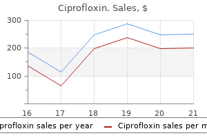
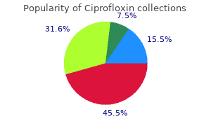
Ciprofloxin
Howard M. Snyder III, MD
- Professor of Urology, University of Pennsylvania School
- of Medicine
- Director of Surgical Teaching, Pediatric
- Urology, Childrenĺs Hospital of Philadelphia, Philadelphia,
- Pennsylvania
Ciprofloxin dosages: 1000 mg, 750 mg, 500 mg, 250 mg
Ciprofloxin packs: 30 pills, 60 pills, 90 pills, 120 pills, 180 pills, 270 pills, 360 pills

Order ciprofloxin 750 mg on-line
Due to the good blood supply in the area of the Papilla duodeni major antibiotic mechanism of action 750 mg ciprofloxin with mastercard, sturdy bleeding might occur virus 85 buy generic ciprofloxin 1000 mg on-line. I Clinical Remarks If gallstones repeatedly lead to the best antibiotics for acne buy ciprofloxin 500mg with visa an inllammation of the gallbladder! This method, the risk of an unintended ligation of the Ductus choledochus with subsequent stasis of the bile (cholestasisl is lowered. Radiography after intravenous utility of contrast agent permits the visualisation of the gallbladder and bile ducts, together with the detection of noncalcified bile concrements. Malignant tumours of the bile ducts (cholangiocarcinoma) or of the pancreas (pancreatic carcinoma! The pancreas is in a secondary retropetltonaal place and initiatives roughly on the primary to second lumbar vertebrae. The head (Caput pancreatisl is adjoining to the Pars descendens of the duodenum and continues as the physique of the pancreas (Corpus pancreatis). A particular feature of the pancreas is that the ache can be projected dorsally onto the same dermatome because of the retroperitoneal place of the organ. The ventral pan- creatic bud folds dorsally (b, e) and fuses, along with the excretory ducts, with the dorsal pancreatic bud (c, fl within the 6111-7111 weelc. The excretory duct of the pancreas is fashioned by the union of the distal dorsal Ductus pancreaticus and the ventral Ductus pancreaticus and enters the Papilla duodeni major. The proximal portion of the dorsal Ductus pancreaticus develops (in 65% of all cases) into the Ductus pancreaticus accessorius which joins the duodenum on the Papilla duodeni m~ nor. In this case, the duodenum has to be minimize and sewn back next to the pancreatic duct system. If the fusion of both pancreatic buds is incomplete (pancreas divlsum), the dorsal Ductus pancreaticus could represent the principle excretory duct (10% of all cases) which may cause repetitive pancreatitis due to a stasis of secretions. In the case of recurring inflam- mation, it needs to be considered whether or not gallstones and alcohol abuse could be dominated out as the trigger. In this case no outflow of pancreatic secretions is feasible via the Papilla duodeni minor. The head of the pancreas (Caput pancreatls) lias on the descending part of the duodenum and has a dorsal uncinate course of (Proc. The subsequent tail of the pancreas (Cauda pancreatisl passes on the dorsal facet of the left colic flexure earlier than the left kidney and extends to the hilum of the spleen. The pancreas has an anterior and a posterior floor (Facies anterior and Facias posterior), that are separated from one another by the boring higher and lower margins (Margo superior and Margo inferior). The anterior facet of the pancreas is covered by parietal peritoneum and varieties the posterior wall of the Bursa omentalis. The posterior side of the pancreas is fused to the original parietal peritoneum of the posterior abdominal wall as a end result of the pancreas was repositioned into the retroperitoneal area throughout its improvement. The exocrine half uses its tail finish (acini) to produce digestive enzymes that are provided as precursors through the system of ducts into the intestinal lumen. Besides other hormones, the islets produce insulin and glucagon which are secreted into the blood and serve to regulate the blood glucose stage. The variation within the opening of the excretory ducts has an influence on the development of pancreatic illnesses. In addition to alcohol abuse, harm to the Papilla duodeni major by gallstones is the most typical explanation for the irritation of the pancreas (pancreatitis), which is caused by a backflow of secretions with autodigestion. A Ductus pancreaticus accessorius with a separate opening can then prove to be helpful when it communicates with the primary duct. Thus its provide is ensured from the drainage areas of the Truncus coeliacus and A. I Clinical Remarks this inteRliNe arterial blood supply via two arteries of the Truncus coeliacus and, as properly as, by the A mesenterica superior clearly explains why infarctions of this very important gland are uncommon. From the inferior margin of the gland, the Nodi lymphoidei pancreatici are linked to the Nodi lymphoidai mesentarici superiores. Due to the retroperitoneal place, there are additionally connections to the retroperitoneal Nodi lymphoidei lumbales. The numerous lymphatic drainage pathways clarify why in circumstances of pancn~~atlc can:lnoma intensive lymph noda metutasn often exist on the time of diagnosis. The parasympathlcus promotes the discharge of the digestive secretions and insulin formation, and the sympathic:us inhibits these capabilities. The sympathetic postganglionic neurons and the parasympathetic pre- ganglionic nerve fibres reach the pancreas from the Plexus coeliacus predominantly via perivascular plexuses. Particularly for the top area, nerve fibres from the lhlncua vagalla polltarlor and infrequently from the Truncus vagalls antertor go on to the gland. Synaptic switching of the parasympathetic fibres is performed by microscopically small ganglia, which are partially embedded within the pancreas. Due to the massive contact area with the diaphragm, the place of the speen is extremely dependent on respiration. Between these two peritoneal duplicatures, the Recessus splenicus of the omental bursa extends as a lot as the hilum of the spleen. The spleen is a secondary lymphatic organ and plays a task within the immune system as properly as in filtering of the blood. The edge facing upwards (Margo superior) between the two areas is often grooved, while the underside edge (Margo inferior) is somewhat clean. I Clinical Remarks Since the spleen sits relatively far cranially in the left epigastrium and initiatives onto rib X. The spleen is surrounded by a fixed capsule from trabeculations of connective tissue within the parenchyma (Pulps splenical. This purple pulp helps with the breakdown of red blood cells (rbc) and the storage of platelets, and is accountable, along with the liver, for the formation of the blood within the foetal period. Stored within the purple pulp there are white nodules that are seen microscopically as white pulp. The course and the branching patterns of the blood vessels are functionally and clinically important: A ´┐Ż. From the trabecular arteries, vessels branch off, surrounded by lymphocytes of the white pulp and accordingly are designated as central artedes. They either terminate as open vessels and the blood flow into the connective tissue mesh of the pink pulp (open circulation! The blood cells, which pass by way of open circulation within the pink pulp, have to enter back into the circulatory system between the endothelial cells within the wall of the sinusoids. From the sinusoids, the blood passes via the pulp veins back into the trabecular veins and thus to the V. At the hilum it divides into two to three main branches after which as a lot as six terminal branches. Here the spleen segments are important: significantly vertical tears, which incorporate multiple segments, bleed closely; conversely, horizontal tears bleed relatively little, since the splenic arteries are functionally terminal arteries. This also explains why infarctions of the spleen often broaden wedgeshaped between the borders of the segments. If the spleen has to be eliminated because of a traumatic rupture, the accessory spleen can take on its operate in order that no immune deficiency occurs. First, the liver has to be mobilised upwards so as to take away the Omentum minus.
Order ciprofloxin 750mg overnight delivery
A tympanostomy tube can then be inserted via the incision for long-term aeration of the middle ear antibiotic and yeast infection buy ciprofloxin 750 mg low price. Tympanic cavity the tympanic cavity (Cavitas tympani) is separated from the ex ternal auditory canal by the eardrum which is airtight and thus represents a closed house which must be ventilated antibiotics xanax generic ciprofloxin 750mg with mastercard. This ventilation of the tympanic cavity (and the aircontaining cells of the mastoid process) solely takes place during the swallowing course of in which the tube antibiotic 5 day discount ciprofloxin 750mg with amex, which is otherwise sealed, opens briefly and permits the air change between nasopharynx and middle ear. The space between Pars flacci da of the tympanic membrane (lateral) and hammer head and an vil body (medial) is the Recessus epitympanicus. The distance between the Epitympanum and the Hypotympanum is 12´┐Ż15 mm at a depth of 3´┐Ż7 mm. The tympanic cavity is lined by a single layer of isoprismatic epi thelium; individual goblet cells and ciliated epithelium are also current. In the Lamina propria there are tubulous glands (Glandulae tympanicae), which are innervated by the Plexus tym panicus. The auditory tube (Tuba auditiva [auditoria]) flows into the tympanic cavity in the decrease wall part. Just be low flows the Canaliculus chordae tympani, through which the Chorda tympani enters the tympanic cavity. It is usually brought on by micro organism and viruses entering the center ear via the pharyngotympanic tube (Tuba auditiva [auditoria]) throughout or after an an infection of the nasopharynx. The irritation is characterised by a reddening of the mucous linings, oedema, granulocytic infiltration and formation of pus. The latter is due to the truth that bacteria (pneumococcal and meningococcal) in the context of an infection of the meninges (meningitis) come via the Aqueductus cochleae (see below) into the inner ear and result in labyrinth failure. If the sound stress transformation fails utterly, the listening to loss lies at roughly 20 dB. Inflammatory foci in the space of the cochlea can also trigger inside ear listening to loss. Between the oval and the round window, the medial tym panic cavity wall bulges out in the course of the Promontorium by way of the basal cochlear coil. Above the oval window, the me dial wall protrudes outward via the lateral semicircular ca nal in the path of the Prominentia canalis semicircularis lateralis. The canal bulges the medial wall in direction of the hor izontally running Prominentia canalis nervi facialis. The Tuba auditiva [auditoria] commences at the Ostium tympani cum tubae auditivae and is demarcated at the top by the Septum canalis musculotubarii from the Semicanalis musculi tensoris tym pani. Vascular, lymphatic and nervous methods Arteries the center ear including the ossicles and the Cellulae mastoideae are supplied by 10 arteries (> Table 9. They are listed right here for the sake of completeness however actual information is superfluous infor Prominentia canalis semicircularis lateralis Antrum mastoideum Prominentia canalis facialis Fenestra vestibuli Canalis nervi facialis Proc. Vertical part in the longitudinal axis of the temporal bone; frontal lateral view. On the entrance of the Corpus is the articular floor for articulation with the Malleus. The base plate is movably fastened within the oval window through a connective tissue ring ligament (Lig anulare stapediale) and transmits the sound vibrations to the perilymph of the inner ear. The three bones are related in a series and linked by true joints (Articulatio incudomallearis, a saddle joint, and Articulo incudostapedialis, a ball joint). Innervation the tympanic cavity, the ossicles and the Cellulae mastoideae are innervated sensitively from the N. A a half of the fibres is switched here in small groups of multipolar ganglion cells to post ganglionic neurons and innervates the tympanic cavity mucosa. Apart from parasympathetic fibres, sympathetic fi bres additionally happen within the Plexus tympanicus (sometimes referred to as R. Low air impedance has to be transferred to the significantly greater internal ear fluid impedance. This requires the amplification of the sound waves (impedance matching), which is achieved by the size difference between the tympanic membrane (55 mm2) and the oval window (3. Its contraction leads to a reduction of excessive sound in tensities and the volume vary is adjusted dynamically. A contraction ideas the stirrup foot plate in the oval window and reduces energy switch. Vascular, lymphatic and nervous techniques the blood supply for the tympanic cavity was already discussed above. Mastoid process In neonates, only the tympanic cavity and the Antrum mastoide um are full of air. First in younger children (finishing approxi mately in the sixth 12 months of life) airfilled cells are shaped within the mas toid course of ranging from the anvil. Zygomatic and temporal bone pyramids may be virtually fully pneumatised (extensive pneumatisation); the pneumatisation may also be com pletely lacking (compact mastoid bone) or solely barely shaped (partially pneumatised). Clinical remarks An inflammation of the Cellulae mastoideae (mastoiditis) is commonly an inflammation transferred from the tympanic cavity. The inflammation can spread from the mastoid to the delicate tissue parts behind and in entrance of the ear, the M. Both branches combine within the depth of the facial canal (Canalis nervi facialis) to the N. It runs in the Os temporale to the entrance medially and exits on the Hiatus nervi petrosi majoris on the Facies anterior of the Pars petrosa ossis temporalis under the dura. The nerve carries preganglionic parasympathetic fibres to the Ganglion pterygopalatinum for the innervation of lacrimal, nasal, palatine and pharyngeal glands and taste fibres from the palate. Ostium pharyngeum tubae auditivae, which protrudes on the aspect to the rear in the nasopharynx (the Ostia of each side protrude as Torus tubarius). The latter consists of a groove manufactured from flexible cartilage (Cartilago tubae auditivae). The upsidedown cartilage groove is medially enclosed by connective tissue (Lamina membra nacea) to form a slitshaped canal. The bony section of the Tuba auditiva [auditoria] is inside a trian gular bony canal (Semicanalis tubae auditivae of the Canalis mus culotubarius) of the Pars petrosa ossis temporalis. Clinical remarks the Tuba auditiva [auditoria] is lined by respiratory ciliated epithelium with goblet cells; combined Glandulae tubariae occur within the subepithelium. Failure of the protective mechanism in the tube can lead to an ascending irritation with formation of a tube catarrh up to otitis media. By injection of air through the nose, adhesions and closures contained in the tubes may be dissolved. One of the most typical causes of conductive hearing loss in childhood is the relocation of the tube opening (tube occlusion) due to tube catarrh or restricted nasal respiratory because of enlarged pharyngeal tonsils (adenoids). If the tube operate dysfunction persists over an extended period of time, restructuring of the middle ear mucosa takes place.
Diseases
- Sulfatidosis juvenile, Austin type
- Anti-plasmin deficiency
- Al Frayh Facharzt Haque syndrome
- Cataract Hutterite type
- Melanoma, malignant
- Placenta neoplasm
Generic 1000mg ciprofloxin mastercard
The veins of the ventricular system antibiotics review discount ciprofloxin 750mg on line, of the basal ganglia infection gone septic buy 500 mg ciprofloxin overnight delivery, and of the inner capsule also belong to the deep cerebral veins antibiotics horses buy discount ciprofloxin 1000mg. After elimination of the cerebellum, the basal cerebral veins are seen, which drain the venous blood from the rhombencephalon, mesence- phalon and insula. The haematoma induced a shift of the centre line; as properly as part of the temporal lobe was pushed by way of the Incisura tentorii underneath the Tentorium cerebelli. Surgery with ligation of the bleeding vessel as soon as possible and relief of the intracranial pressure (by decompression) are essential prognostic elements. In this acute or insidious course of (sometimes taking weeks), venous blood collects between the Dura mater and arachnoid mater, which is associated with unspecific symptoms such as dizziness, headache, fatigue, lack of power or confusion. Subdural haematoma can be associated with intracerebral bleeding and corresponding acute neurological deficits (-+ pp. The therapy consists of a surgical process with insertion of an outflow drainage. Subarachnoid haematoma can often be traced again to ruptu´┐Ż res of aneurysms (pathological bulging of arteries). An early intervention with surgical ligation of the blood source is prognostically essential. Primary grooves (-o Table) as well as Sulci which would possibly be developed and identifiable in each mind, divide the neocortex into 5 lobes (Lobi) seen from the outside. Gyrus precentralis, main somatomotor cortex) are distin- guished from secondary and affiliation fields (a. Although the speech (language) manufacturing is severely restricted, the flexibility to name objects, and the understanding (comprehension) of speech are often preserved. The syntax is usually no longer appropriate and combined with poor articulation of sounds. Unilateral lesions of the first auditory cortex result in impairment of the directional listening to, in addition to to issues within the discrimination of frequencies and intensities. Damage to the prtmary visible cortex on one side leads to cortical blindness with a homonymous hemianopsia. If the secondary cortical areas are affected, the affected person can nonetheless receive visible stimuli, however not interpret and coordinate them (vlau. Lesions in the frontal visible field which may be associated with a failure of the area 8, result in a deviation of the gaze of each eyes to the affected facet (diviation conjugu6e). These areas of the mind are largely unidirectionally related and form a functional unit. As the S-shsped folding of the mediobasal cortex resembles the legendary creature Hippocamp (a kind of sea horse). The archicortex is a part of the limbic system and functionally essential for studying and memory processes. This view from under onto the gyri and sulci of the cerebral hemispheres reveals the Gyrus parahippocsmpalis with the Uncus and the neighbouring Sulcus collateralis. Because it playsloss has great medical an essential position in illnesses with reminiscence. The hippocampus is simply macroscopically seen after opening the inferior hom of the lateral ventricle(. The Fornix is a paired structure composed of Crus, Commissura, Corpus and Columna fornicis. From its origin at the hippocampus and the subiculum in the temporal lobe it varieties an arch above the third ventricle in course of the Corpus mamillare. Before reaching the Corpora mamillaria the two fornices unite (Commissura fomicisl. I Clinical Remarks Neurodegenentive diseases are associated with an insidious destruction of nerve cells. If the neocortex is concerned afterward, the remaining recollections may also be deleted. Fibre connaGiiona exist to the anterior hypothalamic nuclei, the thalamus, and the Habenulae. All constructions proven listed here are part of the limbic system, a useful concept with enter from numerous buildings in the telencephalon, diencephalon, and mesencephalon. The most relevant constructions are each hippocampi, the Corpora amygdaloidea, the Gyri cinguli and the Nuclei septales. The limbic system controls features similar to impulse, studying, reminiscence, feelings, but also the autonomic regulation of meals consumption, digestion, and replica. The Pars anterior connects the olfactory tracts and the olfac- tory cortices of each side. The Pars posterior connects the Paries anteriores of the temporal lobes (particularly the cortex and Corpora amygdaloidea). Visible constructions of the hippocampus are the Digitationes hippocampi of the Pes hippocampi and the Fimbria hippocampi, which transition into the Crus of Fornix. In the rostral path, the Fornix continues by way of the Corpora within the Columnae, which respectively consist of a Pars Iibera and a Pars tecta. Like the Fornix and hippocampus, the Corpora mamillaria are a half of the limbic system. The Gyrus cinguli projects via the Cingulum to the Regio entorhinalis of the Gyrus parahippocampalis. It contains the Bulbus olfactorius, Tractus olfactorius, Nucleus otfactorius anterior. Tuberculum olfactorium, the septal nuclei, the Regio periamygdalaris and Regio prepiriformis. Bulbus and Tractus olfactorius show clear histological variations to the si*layered isocortex. Olfactory cortical areas exert their results on other areas of the mind via connections to the thalamus and the insula. The basal ganglia (nuclei) are concerned in the design of movement sequences, as well as within the regulation of higher brain functions similar to studying, reminiscence, motivation and emotion. The illustration shovvs the topographical relationship between Ventriculus lateralis, Nucleus caudatus, Amygdala, Putamen, Globus pallidus and Thalamus. Many nuclei of the telencephalon are summarised underneath the generic time period basal ganglia. This is characterised by brief tripping steps and a lack of the accompanying arm swings. The impulses coming into t he basal ganglia are processed either in a direct mode, selling motion, or in an oblique mode, inhibiting motion. Damage "to the Nucleus subthalamicua causes issues of t he indirect processing mode. The sufferers perform proximally accentuated, acute catapulting actions (hemiballism) with the unaffected limb on the contralateral side of the physique. Ientum meeencephll Adanohypophyals Neurohypophyals Aqueduclull mesencephall Clatema lnterpeduncularts velum med.
Discount ciprofloxin 250mg otc
Innervation of the tooth Upper jaw the higher jaw enamel are all sensitively innervated by different indi vidual branches of the N antibiotics for uti or bladder infection discount 750 mg ciprofloxin with mastercard. The entire nerve plexus antibiotics for sinus infection z pack purchase ciprofloxin 250mg with amex, innervated by the upper jaw teeth antibiotics for acne philippines buy 250 mg ciprofloxin free shipping, is referred to as Plexus dentalis superior. The higher jaw front teeth are innervated each from the vestibular as well as from the palatine facet. Nerve Innervation space Molars Course On the Tuber maxillae by way of the Foramina alveolaria to the lateral maxillary sinus mucosa In the Canalis infraorbitalis to the maxillary sinus mucosa From the Foramen infraorbitale on the Corpus maxillae to the entrance teeth 1st incisor 9. Lower jaw the lower jaw teeth are solely sensitively provided by a single nerve the N. It passes by way of the Foramen mandibulae within the Canalis mandibu lae, where it forms the Plexus dentalis inferior. The group of masticatory muscle tissue is supported by fur ther muscular tissues of the top and throat area. The Pars superficialis runs obliquely from above downwards in path of the decrease entrance, the Pars profunda runs vertically. It shapes the contour of the rear cheek space and varieties a raphe at the rear fringe of the Ramus man dibulae together with the M. Just above the zygomat ic arch the Fascia temporalisis forms a superficial and a deep layer that are hooked up on the outer and inside floor of the Arcus zygo maticus. The retraction of the alveolar bone can result in problems in the insertion of implants for dental prostheses. It has a smaller upper head (Caput superius) and a bigger lower head (Caput inferius). A part of the muscle fibres of the Ca put superius inserts on the anterior ligament of the Discus articu laris and on the joint capsule of the temporomandibular joint and attaches in bilateral exercise the Caput mandibulae on the tubercu lum slope throughout adduction of the lower jaw. Unilateral exercise results in the grinding movement on the working side and to stabi lising the Caput mandibulae on the dormant aspect. The Caput infe rius is the only masticatory muscle half which is involved within the jaw opening. It induces the oral opening and forces the further mo tion in the type of a mixture of rotation and translation. At the bottom level of the Fossa mandibularis the bone is paper thin and translucent on the cranium. The articular floor passes forward to the vertex of the joint tubercles (Tuberculum articulare). The Tuberculum articulare is lo cated in front of the Fossa mandibularis and varieties a slanting, downward angled articular floor, which is also known as the tubercular slope. On the Tuberculum articulare the cartilage coating is particularly thick, as a end result of here the facility transfer takes place through the Discus articularis. Together with the Fossa mandibularis the Tu berculum articulare forms an Sshaped joint path. Discus articularis the Discus articularis sits like a cap on the joint head and divides the temporomandibular joint into an upper somewhat larger joint (Articulatio discotemporalis) and a decrease joint (Articulatio disco mandibularis). The discus is tightly frontally fused medially and laterally with the joint capsule. It consists of taut collagen fibres and cross es additional to the rear into a extremely vascularised connective tissue, the Plexus retroarticularis. The high layer consists primarily of elastic fibres, which are hooked up to the Fissura tympanosquamosa and the Fissura petrosquamosa. When the mouth is opened, the lower lay er is stretched, while the top layer is relaxed. At the entrance the Discus articularis is connected medially and laterally to the joint capsule. When opening and Discus articularis Fossa articularis Meatus acusticus externus Tuberculum articulare M. Functionally, the temporomandibular joints allow food intake and fragmentation, in addition to articulation when talking and singing. Both temporomandibular joints kind a functional unit and thus work at all times at the identical time. It is formed between the Mandibula and Os temporale from secondary cartilage with a development zone. The joint type is related to the development of the dentition and thus not solely depending on whether or not there are tooth, but in addition on the bite type. Structure the joint head (Caput mandibulae) of the temporomandibular joint types the biconvex curved articular process of the decrease jaw. It articulates with the front a half of the Fossa mandibularis and the Tuberculum articulare of the Os temporale. The axes of the joint heads are slanted and intersect in front of the Foramen magnum at an angle between 150´┐Ż and 165´┐Ż. The articular surfaces of the Caput mandibulae are coated by fibrous cartilage and are primarily on the front of the joint head. Fossa mandibularis the temporomandibular joint pit is positioned on the underside of the Os temporale and is 2 to three times larger than the articular Retro-articular venous pad Proc. Degenerative adjustments (osteoarthritis) are normally related to defects in the lateral area of the Discus articularis (perforation). As the jaw joints are true joints (diarthroses), they can be affected by all ailments, which additionally affect the big limb joints. Stronger violent impacts on the Mandibula can lead to fractures of the Collum mandibulae (Collum fracture). Even without fracture, bleeding typically occurs from the retroarticular venous plexus with difficulties to open the mouth. In older folks, owing to robust atrophy of the bone within the Fossa mandibularis a central fracture of the Fossa mandibularis with an intrusion in the center cranial fossa may happen after falls or knocks on the chin. The pain in all diseases of the temporomandibular joint is commonly projected into the external auditory canal. Biomechanics the temporomandibular joint is a combination joint, which permits for movement in all three planes. Main movements of the jaw joints in the context of mastication are: ´┐Ż Abduction and adduction (opening and closing, elevating and lowering of the lower jaw): this is a mixed gliding hinge motion that takes place bilaterally and symmetrically. The axis of this hinge movement passes via the Foramen mandibularae, which is why the N. For this purpose, deformities of the enamel and occlusion problems can have an result on the motion in the jaw joints. On one side the joint head turns round a vertical axis in the joint socket, whereas the opposite slides ahead on the Tuberculum ar ticulare and separates the tooth on this aspect.
500 mg ciprofloxin
The pharyngeal tonsil enlarges to the extent that the child can now not breathe through the nose virus replication order ciprofloxin 750mg with visa. They have issue concentrating and - if already of faculty age - fall behind at school antibiotic resistance rise generic ciprofloxin 500 mg overnight delivery. In these kids virus 7g7 part 0 generic ciprofloxin 750mg otc, the hyperplastic pharyngeal tonsil has to be surgically eliminated to have the ability to improve the scenario. In addition, a tympanostomy tube is placed right into a decrease quadrant of the tympanic membrane. Middle ear infections are always fraught with threat due to the topographical proximity to neighbouring structures. Q the middls sar has six partitions, through which a middle ear lnflammlltlon may spread to adJacent areas. Already shor11y after taking it Maxi appears significantly improved, the fever lessens, he begins to interact again, to play, and is type of again: to his usual self. In the evening, Maxi as soon as again develops a excessive fever, and the mom administers liquid ibuprofen once more. To be on the protected side, the mother administers liquid ibuprofen once more the following morning. On the third day, the mother takes him back to the kindergarten and asks the nursery teachers to administer the nasal drops to Maxi earlier than he takes a nap at midday. It can lead to perforation of the tympanic membrane, irritation of the mastoid course of (mastoiditis), meningitis, mind abscess, thrombosis of the Sinus sigmoideus. Outer ears that are positioned decrease than that are very generally associated with different (often chromoso- mal) deformities. The exterior auditory canal develops from the posterior part of the primary pharyngeal grocwe, which extends inwards as a coneshaped tube, reaching the entodermal epithelial lining of the tympanic cavity (Recessus tubotympanicus). At the start of the ninth week, the epithelial cells of the acoustic floor proliferate and type an ear canal plate. A persistent plate in the e* tarnal acoustic meatus leads to hereditary deafness. At the start of the fifth week, in the mesenchyme of the first and second pharyngeal arch, the auditory ossicles start to emerge. The malleus and incus emerge from the first pharyngeal arch as derivatives,Ear Development-~ On roughly the 22nc1 day, the floor ectodenn on both sides of the rhombencephalon will thicken. These mobile condensations, termed otic placode, subsequently folds inward, forming an otic pit and then the otic vesicle (otocyst. Each vesicle is divided right into a ventral (rostral) part, from which the saccule as well as the Ductus cochlearis emerge, and a dorul(occlpltal) Medon, from which the Utriculus, semicircular canals and Ductus endolymphati-cus emerge. The ensuing structures are in their entirety referred to as membranous labyllnttl. The external acoustic meatus is shaped by the ectoderm of the primary pharyngeal groove; the entoderm of the first pharyngeal pouch varieties the middle ear. The latter has a very slim connection with the a part of the foregut, which will turn into the nasopharynx. On the lateral tympanic cavity wall, the Recessus tubotympanicus is forming, which grows in course of the advancing pharyngeal groove. The auditory ouicles develop within the mesenchyme from the first and second pharyngeal arch at the beginning of the fifth week. On the outer fringe of the primary pharyngeal groove, six auricular hillocks form firstly of the sixth week, which merge in a fancy method till delivery to form the grownup auricle. This gradually recedes and is changed by an entodermal mucosal lining that covers the whole tympanic cavity. Other causes include infections throughout being pregnant, continual ailments in the mom, medicine, alcohol and nicotine. An inability to hear places severe limits on language studying in addition to structured pondering and communication. Early detection and subsequent therapy early on in life are therefore of immense importance. Presentation of the auricle (Auricula), exterior auditory canal (Meatus acusticus extemusl, tympanic membrane (Membrana tympanica), tympanic cavity (Cavitas tympani), auditory ossicles (Ossicula auditusl. The vibrations are carried as much as the oval window of the internal ear by the auditory ossicles (-. Moreover, the inside ear can also utilise vibrations via the cranium bones (bone conduwon). Wrthin the inside ear, sound vitality continues to journey as a wave (migrating wave). The vestibular organ serves in the notion of rotational and linear accelerations. Movements of the endolymph within the vestibular organ lead to a deflection of cilia of the sensory cells that are synaptically linked with afferent fibres of theN. Apart from the tympanic membrane, the three auditory ossicles are visible in the tympanic cavity (Cavitas tympanij: hammeF-6haped Malleus, anvil-shaped Incus and stirrup-like Stapes, as nicely as parts of the membranous labyrinth (labyrinthus membranaceus, blue). A hedgehog and a bear shut their exterior acoustic meatus in order to not be disturbed by noise throughout hibernation. In order to be succesful of examine the tympanic membrane with an otoscopic mirror or a microscope (otoscopy), the auricle has to be pulled upwards and to the again. This stretches the cartilaginous a part of the external acoustic meatus and the tympanic membrane can be seen (at least partially). Treatment includes topical software of disinfecting agents as properly as native and systemic utility of glucocorticoids and antibiotics. The Pars tympanica of the Os temporals separates the anterior, inferior and posterior elements of the Meatus acusticus extemus. In its superior facet, the bony ring is interrupted by the Incisura tympanica (attachment site of the Pars flaccida of the tympanic membrane). When illuminating the tympanic membrane with its shiny, pearl appearance, a triangular mild reflex in the anterior decrease quadrant is often produced, which permits for conclusions on the stress of tympanic membrane. For ventilation over an extended period of time, a tympanostomy tube (grommet) is inserted by way of the incision. The Pars flaccida of the tympanic membrane is thinner than the Pars tensa and subsequently the preferred location for a purulent middle ear Infection (Otitis media) for a spontaneous perforation. The bitter substances in the cerumen normally serve to keep microorganisms and bugs (flies, small beetles, and so on. The auditory bones of the auditory ossicular chain are linked in a collection and linked by true joints (Art. The auditory ossicles fonn a flexible chain for the transmission of sound waves carried out by the tympanic membrane to the perilymph of the inside ear. Low air impeCaput dance has to be transferred to the considerably greater internal ear air impedance.

Ciprofloxin 750mg cheap
With movements in the joint antibiotics light sensitivity cheap ciprofloxin 1000mg without a prescription, size antibiotics for uti uk buy 250 mg ciprofloxin otc, path and place of the joint resultant will change antibiotic 9 fk unsri order 250mg ciprofloxin mastercard. The actual pressure on the cartilage (intra-articular stress and floor contact) depends not solely on the joint resultant (joint load) but additionally on the size of the surface uncovered to the force. They fill the joint cavity fully and are sometimes hooked up to the joint capsule. They look crescent-like seen from above, and wedge-shaped in profile, and they cowl the margins or the periphery of the joint surfaces (medial meniscus, and lateral meniscus of the knee joint). Joint lips (Labra articularia) these constructions are composed of connective tissue and fibrous cartilage, and they serve to enlarge the sockets of joints. In the human body, joint lips (Labra) are found within the shoulder joint (Labrum glenoidale) and within the hip joint (Labrum acetabuli). Bursae A bursa (Bursa synovialis) is, in principle, structured like a joint capsule. The composition of the fluid is type of similar with the synovial fluid in joints. Clinical remarks If overload, irritation or trauma trigger a bursitis (inflammation of a bursa), the bursa can turn into extraordinarily enlarged, resulting in compression of adjoining buildings. If the bursa communicates with a joint cavity, the inflammation can also have an effect on the joint. Clinical remarks In a Coxa valga (enlarged femoral neck angle), the pressure on the joint will increase and the floor exposed to it decreases. Auxiliary or accessory buildings In a number of joints there are auxiliary or accent intra-articular structures that are essential for the biomechanical function and the vary of movement of joints: ´┐Ż Interposed discs ´┐Ż Disci articulares ´┐Ż Menisci articulares ´┐Ż Joint lips (Labra articularia) In addition, bursae (Bursae synoviales) happen, which kind elastic pads or cushions and allow the sliding of tendons and muscular tissues against bone, in addition to ligaments (Ligamenta) which reinforce the capsule, information or inhibit actions. Intra-articular discs They can compensate for uneven areas (incongruence) of the articulating joint surfaces and are uncovered to compressive forces. Ligaments may be flat or string-like and kind a connection of mobile skeletal parts. If ligaments are integrated into the joint capsule, one speaks of intracapsular ligaments as opposed to extracapsular ligaments, which are separated from the joint capsule by free connective tissue. The skeletal muscles comprise approximately 300 muscular tissues including their tendons and muscle-specific connective tissue. Skeletal muscular tissues exist, other than the axial system, within the tongue, pharynx, larynx, in components of the oesophagus and in the anal area. Each skeletal muscle consists of a various variety of muscle fibres, that are their smallest distinct structural units. Muscles on the again of the thigh (ischiocrural muscles) can, for example, on contraction trigger either knee joint flexion (Punctum fixum and muscle origin match) or within the case of a distal Punctum fixum support a backward tilt of the pelvis within the hip joint. Types of muscle There are a number of methods to grade muscular tissues: ´┐Ż Arrangement of muscle fibres: ´┐Ż parallel course of muscle fibres (to pulling direction of the tendon); in depth movements are potential with little drive ´┐Ż pinnate, i. This known as a tear of muscle fibres or in the case of a extreme damage as muscle rupture (depending on the extent of muscle damage). It is differentiated from muscle pressure by which no macroscopic structural adjustments with destruction of muscle cells and bleeding can be recognised. Muscle ache is a pain that happens after bodily exertion, particularly after high stress of sure muscle elements. It used to be attributed to an acidification of the respective muscle by lactic acid (lactate) but this has been refuted. This is attributable to small microtears in the muscle fibrils with a subsequent inflammatory response. They have the operate to transfer the drive created within the contraction of a muscle to the skeletal components. Types From a structural and practical perspective, a distinction is made between pressure tendons and sliding tendons: ´┐Ż Tension tendons are located in the main path of the muscle and are completely used in tensile stress. On the facet going through the bone/connective tissue construction, pressure is positioned on the tendon underneath strain and slides at this point. Muscle-tendon transition the myotendinous zone is the connection between the muscle fibre and the collagen fibres of the origin and attachment areas. It is characterised by an intensive floor enlargement (factor 10) of the cytoplasmic membrane at the muscle fibre end. In the area of surface enlargement the basal membrane of the muscle fibre is surrounded by a microfibre network (thin collagen fibres). The fibrils of the community interlace internally with microfibres of the tendon and create a set anchorage. Tendon attachment zones Tendon attachment zones are current to adapt the different elasticity modules of connective tissue, cartilage and bone to each other, to avoid detachment or tearing of the tendons within the attachment area. It is attribute that fibrous cartilage is embedded on the insertion website, of which the layer immediately overlaying the bone is mineralised. In the realm of insertion the periosteum is lacking and the collagen fibres penetrate into the bone. The collagen fibres of the planar tendons radiate into the periosteum and anchor the tendon into the cortical bone. Periosteal diaphyseal attachments are characterised by roughness (tuberosities) to the bone. Clinical remarks Overloading of chondreal apophyseal tendon attachment zones can result in degenerative modifications with pain within the space of the attachment zone. In periosteal diaphyseal tendon attachment zones, elevated bone formation with pain generally occurs. Auxiliary equipment for muscular tissues and tendons All muscular tissues and tendons need extra constructions to various degrees, to ´┐Ż adapt to the setting ´┐Ż to defend towards mechanical injury ´┐Ż to stop friction losses and to reduce ´┐Ż energy loss. The fascias enable the muscle to contract virtually invisibly with out the encompassing tissue also contracting. Only in the earlier couple of years have they more and more become the objective of scientific focus. The internal tendon sheath sheet (Stratum synoviale, Pars tendinea) is fused with the tendon, the outer tendon sheath sheet (Stratum synoviale, Pars parietale) with the fibrous layer of the tendon sheath. In the gliding area (Cavitas synovialis) a fluid similar to synovial fluid is emitted. Small blood vessels guarantee the nourishment of the tendon by way of small ligaments of the mesotendon (Vincula brevia and longa) into the tendon. A tenosynovitis stenosans (stenosing tenosynovitis) is attribute of overuse of hand flexor muscular tissues. The overload results in minor accidents in the tendon, which the physique attempts to repair with an inflammatory reaction. The associated swelling of the tendon constricts the tendon sheath (Tenosynovitis stenosans) and leads to the formation of tendon nodules, which for each finger flexion has to move through small circular ligaments (ring ligaments, Ligg. Clinical remarks Injuries of the blood vessels for particular person muscular compartments in the context of main trauma. Retinacula Retinacula are connective tissue retaining ligaments for tissue layers or organs. Tendon sheaths A tendon sheath (Vagina tendinis) encloses a tendon everywhere where it runs immediately on the bone or is deflected (hypomochlion). Tendon Bursa the strain elastic bursae facilitate the sliding of tendons and muscles over bones and tendons (> Chap.
Aconitum carmichaeli (Aconite). Ciprofloxin.
- Dosing considerations for Aconite.
- How does Aconite work?
- What is Aconite?
- Are there safety concerns?
- Nerve pain, feeling of coldness, facial paralysis, joint pain, gout, inflammation, wounds, heart problems, and other conditions.
Source: http://www.rxlist.com/script/main/art.asp?articlekey=96604
Purchase ciprofloxin 750mg line
Palletallymptl nodes: ´┐Ż Nodi lymphoidei parasternales: on each side of the sternum antibiotics for uti augmentin generic ciprofloxin 500 mg amex. They obtain lymph from the anterior thoracic wall antibiotics over the counter 750mg ciprofloxin sale, the mammary gland antibiotics for uti yahoo answers generic ciprofloxin 1000mg line, and the diaphragm. Viaceral lymph nodes with connection to the Trunci brunchomediastinales: ´┐Ż Nodi lyrnphoidei mediastinales anteriores: on both sides of the nice vessels, tributaries from lungs and pleura, diaphragm (Nodi lymphoidei phranici superiores), coronary heart and pericardium (Nodi lymphoidei paricardiaci), and thymus. Nodi lymphoidei mediastinales posteriores: on bronchi and trachea (Nodi lymphoidei tracheobronchiales and paratracheales) and oesophagus [Nodi lymphoidei juxtaoesophsgeales) Lymphatic bunka: the Ductus thoracicus traverses the diaphragm at the entrance of the backbone (. Shortly before draining, it receives the Truncus bronchomediastinalis sinister, which runs independently in the mediastinum, and also the left Truncus subclavius (from the arm) and the Truncus jugularis sinister (from the neck). On the right facet, a brief (1 em) Ductus lymphaticus dexter mostly connects the respective lymphatic trunks and flows within the best venous angle. In this kind of section the proximity of the oesophagus, in the posterior mediastinum. Both constructions are solely separated by the pericardiaI cavity (Cavitas pericardiaca). In transoesophageal echocardiography the spatial proximity of the oesophagus to the guts is used. A depiction of the center and significantly the heart valves using an ultrasound probe inserted into the oesophagus is achieved rather more accurately than through an examination of the outer aspect of the thoracic cage. Medlaatlnum ~ dorsal view; after removal of the posterior thoracic wall together with the backbone. The major lymphatic vessel of the human physique, the Ductus 1horacicus, ascends between the aorta and V. In addition, the Aorta descendens and the azygos system as well as the Truncus sympathicus have been displaced on their passage via the diaphragm. It is fashioned from the lumbar and intestinal trunks under the diaphragm and emerges proper dorsal of the aorta through the Hiatus aorticus. The complete Pars thoracica of the oesophagus and, ventrally from this, of the pericardium and the foundation of the lung (Radix pulmonls) are seen here. The oesophagus emerges via the oesophageal hiatus within the lumbar a part of the diaphragm. It is accompanied by an autonomic nerve plexus (Plexus oesophageus), the parasympathetic parts of which become condensed above the oesophageal hiatus to the vagal bunks. The Truncus vagal is posterior seen right here emerges throughout development, predominantly from the fibres of the best N. The autonomic Plexus pulmonalls is formed significantly strongly dorsally and accompanies the principle bronchi to the hilum of the lung. Most difficult to identify is the quick Ductus lymphaticus dexter, which drains into the proper venous angle (between the V. Before reaching its outlet it receives the Truncus bronchomediastinalis, the Truncus subclavius and the Truncus jugularis (not shown). A distinction is made between a systemic cln:ullltlon I= giant circulation) and a pulmonary cin:ullltion (=small circulation), which are linked in a sequence. The heart is the father or mother organ of the cardiovascular system and drives the circulation as a suction and pressure pump. Accordingly, the heart is divided into two halves, which each include an atrium and a ventricle (Ventriculus). In this fashion, blood is pumped from the center into arteries and returned via veins again to the heart. Oxygen content for this division is negligible I In the systemic circulation, oxygenated blood from the left ventricle (Ventriculus sinister) is directed by way of the primary artery (aorta) and downstream arteries into outlying areas, where the oxygen is used and carbon dioxide absorbed. The venous blood is correspondingly deoxygenated and is returned to the heart through the veins, which join to the sucavae superior and inferior) previous to perior and inferior venae cavae entering the right atrium (Atrium dextrum). This blood is then pumped from the best ventricle (Ventriculus dexter) via the Truncus pulmonalis and the pulmonary arteries into the pulmonary circulation, the place renewed oxygen absorption into the blood and exhalation take place. The pulmonary veins transport the oxygenated blood back into the left atrium (Atrium sinistrum) in order that the circulation is completed. As the heart circulates blood around the body, its functions are equivalent to those of blood. The most essential features of the cardiovascular system are: ´┐Ż oxygen and nutrient supply of the organism (transport of respiratory gases and nutrients) ´┐Ż thermal regulation (heat transfer in blood ´┐Ż defence perform (transport of immune cells and antibodies) ´┐Ż hormonal management (transport of hormones) ´┐Ż haemostasis (transport of blood platelets and coagulation factors) the center is divided right into a left and rfght half by the cardiac septum. The two halves of the guts are every subdivided by valves (atrioventricular valves) right into a right and left atrium and a proper and left ventricle. Pars membranacea, whereas the largest part consists of cardiac muscle (Pars muscularis). Within the periphery of the body and the organs the vessels of microcirculation are related between aFteries and veins. Here blood strain is reduced in the arterioles so that oxygen and gas change can happen within the capillaries. The gentle margin of the heart projects from the third to sixth costal cartilage in a line which is 2 em lateral to the proper sternal border. Thellrft margin of the heart projects onto a connecting line between the lower rim of the 3 111 rib (2-J em parasternal) and the midclavicular line on the left. Its size corresponds to the fist of every respective person; the burden is on common 25o-. The heart has four aides: the ventrally oriented Facies stemocostalis corresponds predominantly to the right ventricle. The caudally pointing Facies dlaphragmatlc:a consists of elements from both ventricles. The Facies pulmonalia is fashioned to the best by the best atrium and to the left predominantly by the left ventricle. Since the atria were seen for a protracted time as a part of the upstream veins, naming the posterior side was apparently distributed with. The largest a half of the Facies sternocostalis is covered on both sides by lung and pleura. These areas correspond to the Recessus costamediaatlnales of the Cavitas pleuralis. Underneath the 41h rib, the pleural edges transfer other than each other and confine between them the Trigonum pericardiacum, in which the pericardium lies directly against the ventral thoracic wall. Tapping of the heart lpen:ussion) may give an initial indication of the dimensions of the center. The projection of the guts contours, that are covered by the pleura of the costomediastinal recess. When this subject extends to the left over the midclavicular line, it is an indication of left ventrieuler hypertrophy. However, this measure is comparatively risky and has due to this fact largely been abandoned. In most circumstances an enlargement to the left aspect (left pulmonary surface) is current, which factors to damage to the left ventricle.
Order ciprofloxin 750 mg otc
In the case of intensive pneumatisation the bony partitions are often only extremely skinny antibiotic for ear infection purchase ciprofloxin 250mg without prescription. Clinical remarks Inflammation of the paranasal sinuses (sinusitis) is a standard illness virus quotes buy discount ciprofloxin 500 mg on line. In kids the ethmoidal sinuses are particularly affected antibiotics for uti how long does it take to work purchase 250mg ciprofloxin otc, in adults the maxillary sinus, but in addition frontal and sphenoidal sinuses can turn out to be infected. Unilateral Sinus maxillaris inflammation is usually brought on odontogenically (odontogenic Sinusitis maxillaris). The inflammation usually originates at the 2nd premolar or the 1st molar (see above). Feared problems of ethmoiditis are the spreading to the Orbita via the thin Lamina papyracea (orbital phlegmon) and the spreading of the irritation through the bony walls of the Canalis opticus to the N. If the Sinus frontalis is highly developed occipitally by way of the orbital roof (Recessus supraorbitalis), clinicians discuss with a dangerous frontal sinus. A frontal sinus irritation can lead via the skinny bony partitions of the anterior cranial fossa. Malignant tumours of the nostril have turn out to be less frequent due to improved occupational well being and security measures. Both arteries penetrate via the corresponding Foramina ethmoidalia anterior and posterior into the ethmoidal complex and run by way of the Canales ethmoid ales between the ethmoidal cells up into the nasal cavity. It passes via the Fossa pterygopalatina through the Foramen sphenopalatinum into the nose and splits into the Aa. The arterial blood supply of the paranasal sinuses is carried out for: ´┐Ż the maxillary sinus via a department of the A. The nasal cavities lead the blood into the Plexus cavernosi concharum and different venous networks of the nasal mucosa. The paranasal sinuses drain their blood in several ways: ´┐Ż the maxillary sinus in vascular networks of the dental roots to the Plexus pterygoideus ´┐Ż the ethmoidal cells in Vv. Lymph vessels the Nodi lymphoidei submandibulares are regional lymph nodes of the external nose and nasal atrium. The lymph of the nasal cavi ties and paranasal sinuses is drained largely in the course of the throat to the Nodi lymphoidei retropharyngeales and from here to the Nodi lymphoidei cervicales profundi and to a lesser extent ahead to the Nodi lymphoidei submandibulares. Innervation the nostril and the paranasal sinuses are innervated sensitively by branches of the N. The innervation of secretory lively glands and of the blood vessels in the nostril and paranasal sinus mucosa is supplied through parasympa thetic fibres of the N. Sense organ innervation: ´┐Ż From the olfactory fields, the Fila olfactoria passes by way of the Lamina cribrosa of the cranium base. After switching within the Bulbus olfactorius, the fibres proceed via the Tractus olfactorius to the central core areas. After switching, the postgangli onic fibres run together with the sensory fibres of the N. Sympathetic innervation: ´┐Ż Postganglionic fibres from the Ganglion cervicale superius cross as N. Arnold Skills After working through this chapter, you want to be in a position to: ´┐Ż name all oral structures ´┐Ż describe the nerve course in the oral cavity ´┐Ż clarify the vascular provide to the oral cavity ´┐Ż explain the development of the enamel ´┐Ż describe the detailed construction of the various tooth ´┐Ż describe the structure and performance of the Articulatio temporomandibularis, and the situation and function of the masticatory muscle tissue ´┐Ż describe the construction, location and capabilities of the tongue and of the palate ´┐Ż reproduce the situation, construction and performance of the salivary glands ´┐Ż clarify the construction of the floor of the mouth and its compartments ´┐Ż name the blood supply, innervation and lymph drainage of the abovementioned structures and organs ´┐Ż describe the topographical location and the neighbourhood relationships of the buildings and organs to each other and to be capable of classify the encircling areas and describe their operate ´┐Ż explain the development of the oral cavity, masticatory apparatus, tongue, palate and salivary glands Clinical remarks A hyposmia (decreased olfactory sensation) or an anosmia (missing olfactory sensation) could be brought on by viral infections, chronic sinusitis, obstructions because of relocation of the airways to the olfactory mucosa. Clinical case Perimandibular abscess Case study A 50-year-old male affected person, who underwent root canal treatment 1 12 months ago due to profound caries of tooth 46, presents with considerably lowered general health and an elevated temperature of 38. Initial examination the pulse rate of the patient is increased, swallowing difficulties and limited mouth opening are clear. The medical data present that within the preceding investigations the affected person has had gingival recession (inflammation-free gum recession) and periodontal pocket depths as much as 12 mm. The proper cheek and the throat space at the proper under the decrease jaw are clearly swollen, whereby the swelling is simply native and consists of the submandibular delicate tissue. Clinically, this gives the impression of a selection of the swelling into the parapharyngeal area. Diagnostics From a differential diagnostic point of view the medical findings indicate a perimandibular abscess on the best aspect, a salivary calculus in the Glandula submandibularis or a tumour of the Glandula submandibularis. In order to rule out a salivary calculus or tumour, an ultrasound examination is conducted. Treatment the dentist opts for the intraoral opening of the abscess and the administration of an antibiotic. After opening the abscess, the dentist inserts a drainage, so pus and wound secretions can be drained away. After therapeutic the renovation of the root filling follows with apicoectomy in a second session. In addition, he tells the patient to come every quarter yr to check the periodontal situation with professional tooth cleaning. In the oral cavity the ex cretory ducts of three large salivary glands emerge (Glandula pa rotidea, Glandula submandibularis and Glandula sublingualis), their secretions form the total saliva, which moistens the oral cavi ty. Development In evolutionary phrases, the event of the oral cavity originates from 2 germ leaves. The rear part evolves from the entodermal foregut, the front part from the ectodermal mouth recess. Through the development of the entrance brain the unpaired brow bulge grows ahead and downwards, whereas the 2 paired maxillary and mandibular bulges develop forwards. The major oral cavity is a uniform oral nasal area which is separated from the foregut by the pharangeal membrane (Membrana buccopharyngea). As early as the 3rd embryonic week the buccopha ryngeal membrane tears, in order that the Stomatodeum and the intesti nal tube are connected to each other. Approximately in the 6th em bryonic week the first palate types from the forehead bulge of the unpaired main palate; from the 2 maxillary bulges the palatinal processes emerge laterally. These develop medially in the direction of each other, where they fuse in the midline, in order that oral and nasal cavity are completely separated from each other. Frenulum labii superioris Uvula palatina Palatum durum, Raphe palati Palatum molle [Velum palatinum] Arcus palatopharyngeus M. Laterally, the oral cavity is restricted by the cheeks, the muscular base is the M. The roof of the oral cavity varieties the palate, which is divided into the onerous palate (Palatum durum) and the taste bud (Palatum molle). Orientation Principally, the directional terms of the body additionally apply to the oral cavity: nonetheless, since the teeth are arranged in a ellipsoid dental arch, extra directional names are required. Dental surfaces, which face the Vestibulum oris are within the entrance tooth region in both the higher and decrease jaw the labial surfaces, in the posterior area the vestibular and buccal surfaces. The surfaces going through the oral cavity are referred to within the decrease jaw as lingual, within the higher jaw as palatinal surfaces. Tooth surfaces that are in the direction of the throat, are referred to as distal, tooth surfaces within the direction of the Rima oris, as mesial surfaces.
Buy cheap ciprofloxin 750mg online
Children usually have a tendency to antibiotics for dogs after surgery generic ciprofloxin 500mg amex have astrocytomas whereas adults are more doubtless to bacteria are the simplest single cells that discount ciprofloxin 500 mg on line have metastatic spinal tumors virus 4 pics 1 word discount 1000 mg ciprofloxin fast delivery. It is estimated that 5´┐Ż10% of most cancers sufferers will develop symptomatic spinal metastasis. The backbone is the third commonest website for cancer cells to metastasize (following lung and liver). Elderly sufferers have an elevated frequency of spinal meningiomas with a female-tomale ratio of 4:1. Disorder Description: Extramedullary tumors include neurofibromas, meningiomas, schwannomas, and 616 Spinal Perimedullary Fistula metastatic tumors. Metastatic spinal tumors are commonly due to prostate, breast, and lung cancers, lymphomas, and leukemias. Intradural intramedullary spinal tumors embrace ependymomas, which are the commonest kind of intramedullary tumor in adults (50´┐Ż60%), and gliomas, which are comprised of astrocytomas and high-grade gliomas. Symptoms can happen after direct compression, ischemia as a result of vascular infiltration, or invasive infiltration. In addition, due to their affiliation with urinary retention from spinal wire compression, patients are at higher risk of urinary tract infections and pyelonephritis. Treatment Complications: Chemotherapy, radiation, radiosurgery, and radical resection may be considered relying on the type and placement of the tumor. Radiation, radiosurgery, and surgery dangers include paralysis, bladder incontinence, and urinary retention. Risks of chemotherapy include infection, hair loss, and gastrointestinal discomfort in addition to multiple different systemic unwanted effects dependent on the treatment. Symptoms Localization web site Spinal cord Comment Pain and spinal tenderness usually precede neurologic deficits. Imbalance with strolling occurs when dorsal columns are affected May be concerned in the instances of intradural or intramedullary tumors. Spinal Perimedullary Fistula Epidemiology and Demographics: Most generally these are small fistulae and happen in older adults. Less generally, medium-sized fistulae could have an result on young adults and rarely large fistulae may affect kids. Disorder Description: Abnormal connection between a spinal artery and medullary vein. Symptoms often develop slowly over time but the childhood giant fistulae may present abruptly and even as subarachnoid hemorrhage. Anterior horn cells Conus medullaris Cauda equina Specific spinal roots Symptoms Localization site Spinal wire Comments Gradual myelopathy consisting of weakness and numbness under the level of the lesion. Progressive urinary and fecal incontinence can be seen the young adult kind is usually located here and presents with paraplegia and outstanding incontinence Syndromes with mixed higher and lower motor neuron deficits Conus medullaris 617 Section 1 Diagnostics Secondary Complications: Some varieties are associated with vascular malformations within the pores and skin and mind. Spondylitis Epidemiology and Demographics: Incidence is approxiDisorder Description: Spondylitis is defined as inflammation of the vertebrae. Spinal Subarachnoid Hemorrhage Epidemiology and Demographics: this disorder is extremely rare with few circumstances reported in the literature. Disorder Description: Hemorrhage accumulating between the arachnoid and pia mater within the spinal twine. This is usually related to trauma or spinal procedures but may additionally be seen in coagulopathy. Spinal aneurysms are hardly ever present, with or with out spinal perimedullary arteriovenous fistulae. Being within the subarachnoid area, the blood irritates the spinal nerve roots rather than inflicting compression of the spinal twine itself. Symptoms Localization website Cervical spine Comment Neck pain, radicular symptoms together with radiation into shoulders, decreased vary of movement Mid again pain Low back pain, radicular symptoms, hip ache, buttock ache Temporal mandibular joint ache Iritis or anterior uveitis Thoracic backbone Lumbosacral spine Jaw Symptoms Localization web site Cauda equina Comment If the blood collected below L1, then the cauda equina shall be compressed causing ache with numbness and weakness affecting all lumbosacral roots If the blood collects proximally, it may irritate particular person nerve roots causing apparent radiculopathy Eyes Secondary Complications: Cauda equina syndrome Specific spinal roots can happen in sufferers with longstanding spondylitis. Urinary retention and/or incontinence, lack of bowel management, sexual dysfunction, and weakness of the lower extremities should raise the potential of cauda equina syndrome. Ankylosing spondylitis and related spondyloarthropathies: the dramatic advances prior to now decade. Time tendencies in incidence, clinical options and cardiovascular disease in ankylosing spondylitis over 3 a long time: a population based mostly research. Treatment Complications: Most instances are treated surgically with the attendant risks of bleeding, an infection, anesthesia, and harm to underlying tissue. Worse in immunocompromised, together with diabetes, alcoholism, and continual obstructive pulmonary illness. Transmitted through soil or different organic materials contacting the skin, or via inhalation. Staphylococcus aureus Epidemiology and Demographics: Ubiquitous bacterium Disorder Description: Causes quite so much of neurologic that colonizes the pores and skin of many individuals. Infective endocarditis can lead to embolic strokes and mycotic aneurysm; pyomyositis, an infection of the skeletal muscle, is possible. Meningitis happens within the setting of head trauma or neurosurgery (especially shunts) and infrequently due to bacteremia. Symptoms Localization web site Cerebral hemispheres Mental status and psychiatric aspects/complications Brainstem Cerebellum Cranial nerves Comment Meningitis Encephalopathy because of meningitis Meningitis Meningitis Meningitis Symptoms Localization site Cerebral hemispheres Mental status and psychiatric aspects/complications Brainstem Cerebellum Cranial nerves Comment Meningitis Confusion, lethargy, delirium, coma Meningitis Meningitis Meningitis If infection is due to lumbar puncture Infection associated to gadgets and procedures Related to inflammatory state Critical sickness polyneuropathy Direct infection Secondary Complications: Skin ulceration and secondary Conus medullaris Cauda equina Plexus Peripheral neuropathy Muscle an infection by different organisms. Treatment Complications: Treatment with itraconazole or amphotericin B (nephrotoxicity) in additional extreme circumstances, together with meningitis. The upside of bias: a case of continual meningitis as a result of Sporothrix schenckii in an immunocompetent host. Secondary Complications: Lungs, spleen, joints, bones, and/or skin can all be concerned. Treatment Complications: Resistant strains make polytherapy necessary and, with it are more unwanted effects of antibiotics. The incidence in Europe is decrease (10´┐Ż16/100,000 population) compared with the United States (18´┐Ż 41/100,000 population). American ethnic minorities have a higher incidence (57/100,000) than whites (20/100,000). It is a condition which might have longterm consequences (after time level t2), including neuronal death, neuronal injury, and alteration of neuronal networks, depending on the kind and length of seizures. The commonest etiologies embrace infections, brain injury and epilepsy, withdrawal or adjustments in antiepileptic drugs, in addition to remote symptomatic epilepsy. Autoimmune encephalitis has been recently acknowledged as an important etiologic consideration. If sufferers fail to reply, intubation and use of steady infusion of sedatives corresponding to propofol, midazolam, or pentobarbital are incessantly employed. Focal seizures, which can include delicate scientific motor findings similar to facial, eyelid, or limb clonus. Findings range with the type of seizures, seizure severity, period, underlying etiology, and chronologic age of the affected person Mental standing and psychiatric aspects/ problems Secondary Complications: Common complications include respiratory acidosis with or with out metabolic acidosis, hypoxia, hyperglycemia and peripheral leukocytosis, arrhythmias, cardiac troponin elevation, and ischemic electrocardiographic patterns. Rhabdomyolysis may end in acute renal failure, myoglobinuria, transaminitis, and disseminated intravascular coagulation.
Purchase ciprofloxin 750mg on line
In entrance antibiotic injections 500mg ciprofloxin overnight delivery, the labia minora cross with a ribbon of tissue (Frenulum clitoridis) to the glans of the clitoris (Glans clitoridis) antibiotics for uti male generic ciprofloxin 1000 mg on line. The two cavernous bodies (Corpora cavemosa clitoridis) kind a brief physique (Corpus clitoridis) antibiotic bone cement buy ciprofloxin 750 mg visa, which caudally ends with the glans before separating into the Crura clitoridis which are anchored to the inferior ischiopubic rami. Developmentally there exist soma similarities between the construction of the clitoris, which also has a prepuce (Preputium clitoridis), and the structure of the penis. Also the filling mechanisms of the erectile tissue and erection are related in each sexes. The first half in the development of the exterior genitalia is equivalent in each sexes (indifferent gonad). The anterior wall of the Sinus urogenitalis indents to form the urethral groove which is bordered on each side by the urathral folds. To the lateral of these are thelabioserotll folds and anterior is the genital tubercle. Functionally, the ovary serves to mature follicles (and ovulation) and to produce feminine sex hormones loastrogens and progesterone). They transport the ovum into the uterus, the place the event of the kid takes place during being pregnant. In the female, the primordium of the primitive gonad then develops into the ovary. Similar to the testis, the ovary also develops in the lumbar region on the degree of the mesonephros. The ovary nestles into an indentation, which is created by the branching of the A. The shut topographical relationships between the adnexa (ovary and Tuba uterine) and the vermiform appendix (Appendix vermiformisl of the colon e>cplain why inflammations of the appendix (appendicitis) as nicely as those of the uterine tube salpingitis! Between the uterus and urinary bladder is the Excavatio vesicouterina as an enlargement of the stomach cavity. Internally the wall of the uterus is made from a mucous membrane (endomebium), which structure and thickening change through the feminine menstrual cycle, in order to allow the implantation after fertilisation of an ovum. It is connected to a thick layer of anooth musc:l´┐Ż (myomllllluml, of which the muscle fibres present totally different preparations. Parts of ligaments are referred to in accordance with the nomenclature of the layers of the wall. The area inside the uterus is divided into the Cavitas uteri in the physique and the Canalis cervicis uteri within the Cervix of the uterus. The Vllgina is a hollow muscular organ of about 10 em in length in a subperitoneal location. At the inside floor, both the anterior and posterior walls of the vagina (Paries anterior and Paries posterior) reveal transverse mucosal folds (Rugae vaginalesJ. The frontal part additionally exhibits the construction of the uterine wall: the internal mucosal layer (Tunica mucosa, endometrium), then the strong muscular layer (Tunica muscularis, myometrium) of clean muscles, and the outermost peritoneal lining (Tunica serosa, perimetrium). The stroma of the ovary incorporates the follicles (Folliculi ovarici), which contain the egg cells, and later in the feminine cycle turns into the yellow body referred to as Corpus lutaum. It is split into a physique (Corpus uteri) with a superior fundus (Fundus uteri) and a neck (Cervix uteri) which are distinct from one another by a constriction (Isthmus uteri). The uterine tubes are connected to the physique of the uterus (Tuba uterina) on both sides offering a connection to the ovary. The utBrina tube (Tuba uterina [Salpinx]) is 10-14 em long and has several components: ´┐Ż Infundibulum b. Uterus, uterine tubes and (N&ry are inbaperitonaal and therefore have their own peritoneal dupllcatu. The bend is essential for fixing the uterus in place, as a result of it stabilises it in its anteversion in relation to the vagina and hence prevents the uterus from prolapsing on account of a rise in intra-abdominal pressure, for instance because of coughing and sneezing(. The connective tissue within the lesser pelvis is divided from the scientific point of view in the neighborhood of the person organs; particular person fibres are referred to as bends, although such a distinction in anatomical terms is clearly unimaginable. The superior part (Portio supravaginalis) goes through a constriction Isthmus) on the inside cervical os (Ostium anatomicum uteri internuml into the cavity of the uterus Cavitas uteri). The vagina has anterior and posterior partitions (Parietes anterior and posterior), both revealing transverse folds (Rugae vaginalesl. It culminates within the vestibule Vestibulum vaginae), which is already regarded as one of many eJto tarnal genitalia. At the cranial finish, the Portio vaginalis of the cervix surrounds the vagina with the vaginal vault (Fornix vaginae). This place acts as safety and prevents the uterus from bulging out by way of the vagina as a result of a rise in intraabdominal pressure (sneezing, coughing). The fimbriae, which are organized round this opening, are in contact with the surface of the r:Nary and receive the bouncy ovum throughout ovulation. Most different photographs in textbooks are primarily from dissections of elderly girls, in whom the ovaries and uterus are often atrophied. The tubal patency may be checl<ed with an X-ray distinction imaging of uterus and tubes (hystaroealplngography), to be able to rule out stenosis of the uterine tubes within the case of infertility, which for instance may result from inflammation. Today this procedure is often earned out using ultrasound imaging in which a distinction agent is also used. K = portio adapter of the injection probe for the contrast agent 294 Retaining Bands and Connective Tissue Area. The examination should be carried out a minimal of annually for early detection and elimination of any degeneration as a precursor of 8 malignant tumour (cervical cancer). Cervical cancer is among the commonest malignant tumours in girls underneath the age of forty. Since the rectouterine house represents the lowest part of the peritoneal cavity, in upright sufferers it may be a focus point for inflammation of the peritoneum (peritonitis) or the unfold of tumour cells. The creating child within the uterus is nourished by way of the placenta which develops from maternal and foetal tissues after implantation. Up to parturition, the uterine volume will increase 800-1,200 times and the utefine weight increases from 30-120 g to 1,000-1,500 g. After the gas and nutrient trade, the blood is fed again through a umbilicalis) to the child. On the masingle vein in the umbilical wire the mal facet, the placenta is anchored in the mucous lining of the uterus. It can be seen in the image that the placenta is split into 10-40 practical fold´┐Ż (cotyleclona). After the birth, it should be ensured by inspection that the placenta is complete, as any placental residues remaining within the uterus can lead to severe bleeding and infection. The Plexus hypogastricus inferior and Plexus utarovaginalis contain sympathetic (green) and parasympathetic (purple) nerve fibres. The (mostly) postganglionic sympathetic fibres to the ovary move through the Plexus ovaricus alongside the A. Preganglionic peruympathatlc nerve fibres cross from the sacral parasympathicus (S2-S4) through the Nn.
References
- Biedler JL, Riehm H. Cellular resistance to actinomycin D in Chinese hamster cells in vitro: cross-resistance, radioautographic, and cytogenetic studies. Cancer Res 1970;30(4):1174-1184.
- Cruz D, Bagshaw S, Maisel A, et al. Use of biomarkers to assess prognosis and guide management of patients with acute kidney injury. Contrib Nephrol. 2013;182:45-64.
- Bossone E, Rampoldi V, Nienaber CA, et al: Usefulness of pulse deficit to predict in-hospital complications and mortality in patients with acute type A aortic dissection, Am J Cardiol 89:851, 2002.
- Columbus. The Columbus Investigators: Low-molecular- weight heparin in the treatment of patients with venous thromboembolism. N Engl J Med 1997;337:657-662.
- Kane CJ, Bolton DM, Connolly JA, et al: Voiding dysfunction in human immunodeficiency virus infections, J Urol 155(2):523n526, 1996.
- Hirst AE, Gore I. The etiology and pathology of aortic regurgitation. In: Doroghazi RM, Slater EE, eds. Aortic Dissection. New York: McGraw-Hill Book; 1983:13.
- Matsui Y, Utsunomiya N, Ichioka K: Risk factors for subsequent development of bladder cancer after primary transitional cell carcinoma of the upper urinary tract, Urology 54:279n283, 2005.
- Abdel-Fattah, M., Ramsey, I., Pringle S, Hardwick C, Ali H., Evaluation of transobturator tapes (E-TOT) study: randomised prospective single-blinded study comparing inside-out vs. outside-in transobturator tapes in management of urodynamic stress incontinence: Short term outcomes. Eur J Obstet Gynecol Reprod Biol 2010;149:106-111.